Figure 7.
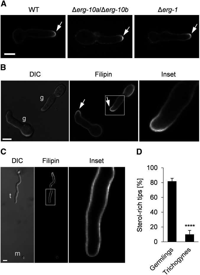
Membrane sterol distribution differs between vegetative and sexual cells of N. crassa. Germlings and trichogynes were stained with filipin. (A) Accumulation of sterols at the polarized cell tips of WT (FGSC 2489), Δerg-10a/Δerg-10b (N4-38), and Δerg-1 (MW_308) germlings. (B) Arrows indicate sterol-rich domains in interacting WT germlings (g). The white square marks the area of the enlarged image to the right. (C) Trichogyne (t)–microconidium (m) interaction. The white rectangle indicates the position of the enlarged image to the right. Bar in (A–C), 5 μm. (D) Quantification of sterol-rich cell tips in germlings and trichogynes during cell-to-cell interactions. Values represent the mean ± SD from three and two independent experiments with ∼100 germling pairs and up to 40 trichogynes per replicate (****P < 0.0001, unpaired, two-tailed Student’s t-test). FGSC, Fungal Genetics Stock Center; WT, wild-type.

