Figure 1.

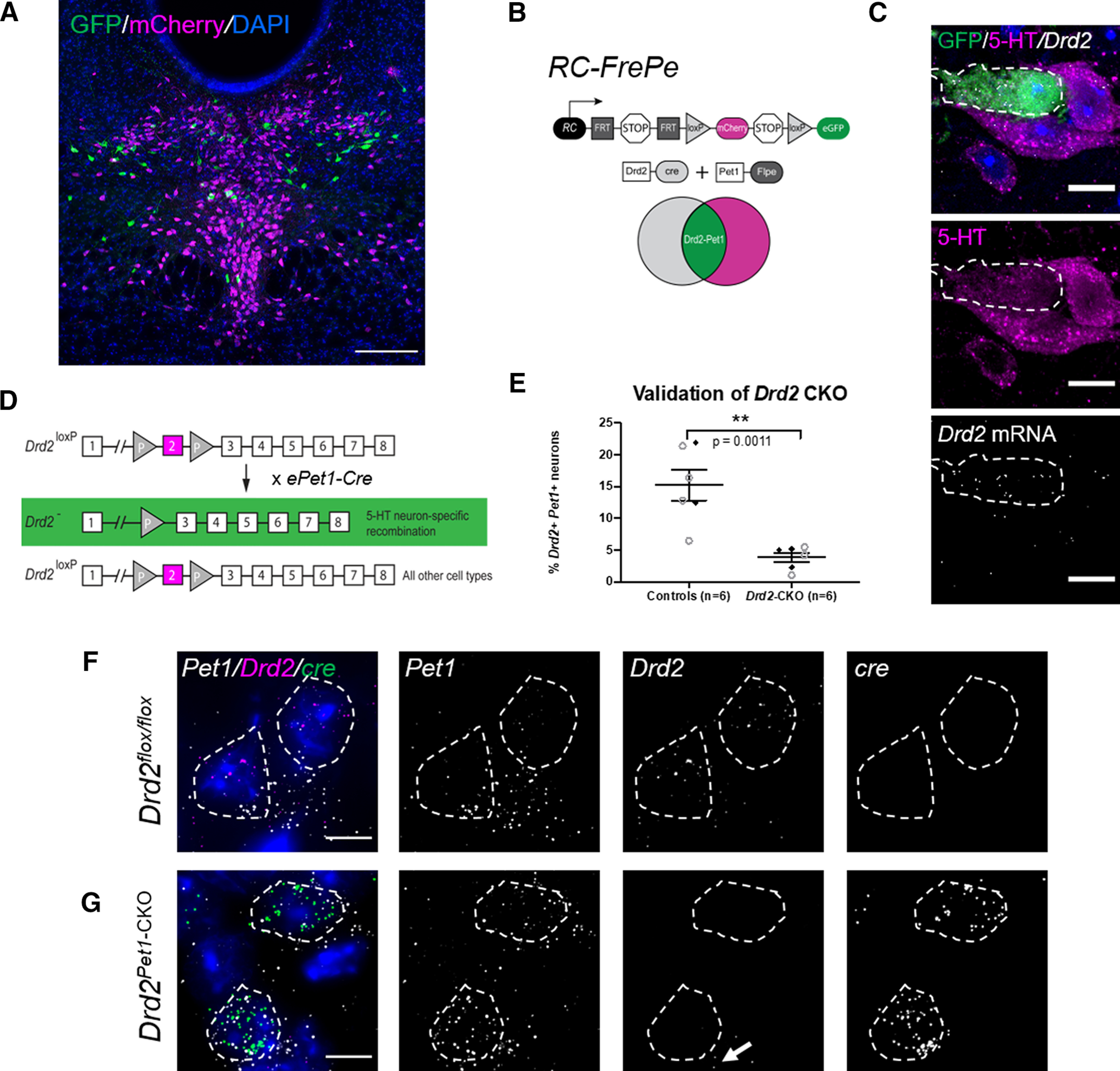
Visualization of Drd2-Pet1 serotonergic neurons and the loss of Drd2 gene expression in Drd2Pet1-CKO mice. A, Drd2-Pet1 neurons are intersectionally labeled with GFP (green) and Pet1-only positive cell bodies labeled with mCherry (magenta) in a coronal brain section of the DR from a P90 triple transgenic Drd2-Cre;Pet1-Flpe;RC:FrePe mouse. Scale bars: 200 μm. B, Intersectional genetic strategy: expression of Drd2-Cre and Pet1-Flpe transgenes results in dual recombination of intersectional allele, RC:FrePe, labeling cells expressing Drd2 and Pet1 with GFP. C, Dual immunohistochemistry for GFP (green) and 5-HT (serotonin, magenta) coupled with FISH detection of Drd2 mRNA, which shows co-localization of intersectionally labeled Drd2-Pet1 neuron cell bodies with 5-HT and Drd2 mRNA. Scale bars: 10 μm. D, Strategy for conditional deletion of Drd2 in serotonergic neurons (referred to throughout as Drd2Pet1-CKO). Cre recombination excises Drd2 exon 2 (magenta) producing serotonergic-specific (boxed in green) deletion of Drd2 gene sequences. E, Percentage (mean ± SEM) of Pet1+ serotonergic neurons that express Drd2 in control (n = 6) versus Drd2Pet1-CKO (n = 6) shows reduction of Drd2 expression in Pet1+ neurons (controls: 15.23 ± 2.41 Drd2-Pet1 dual positive neurons per brain, Drd2Pet1-CKO: 3.87 ± 0.73 Drd2-Pet1 dual positive neurons per brain, p = 0.0011, unpaired t test). Filled black diamonds represent male mice, open gray circles represent female mice. F, G, FISH on (F) control and (G) Drd2Pet1-CKO tissue. Drd2 transcripts detected in Pet1+ cells in control sections, but not in Drd2Pet1-CKO mice, indicative of loss of Drd2. cre transcript is not present in control (F, far right) but is present in Drd2Pet1-CKO Pet1 cells, as expected (G, far right). Pet1, Drd2, and cre transcript are shown separately in grayscale. Note Drd2 expression remains in non-Pet1 cells (arrow). Dotted lines drawn to encircle DAPI nuclei. Scale bars: 25 μm.
