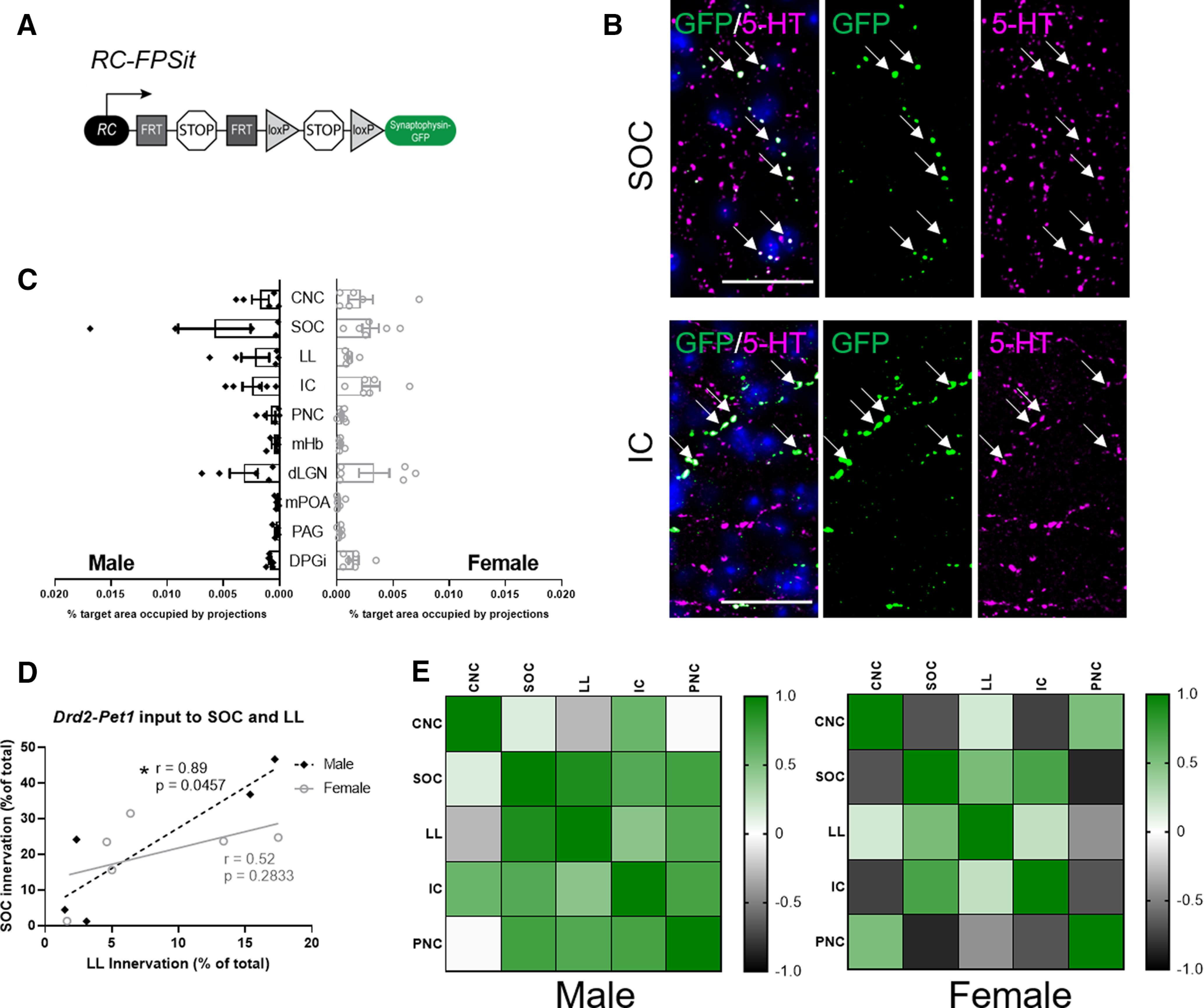
Figure 9.

Drd2-Pet1 neuron axon terminals target brain regions involved in sensory processing and defensive behavior in both male and female mice. A, Intersectional genetic strategy: expression of Drd2-Cre and Pet1-Flpe transgenes results in dual recombination of intersectional allele, RC-FPSit, to label boutons of Drd2-Pet1 neurons with Synaptophysin-GFP. B, Representative images of Drd2-Pet1 boutons in the SOC and IC. GFP+ (green, marked with arrows) boutons co-localize with 5-HT (magenta) staining. DAPI-stained nuclei shown in blue. Scale bar: 25 μm. C, Quantification of the percent target area occupied by projections for all ten brain regions examined (for quantification protocol, see Materials and Methods). Target areas analyzed include brain regions involved in auditory processing and social behavior including the CNC, SOC, LL, IC, PNC, mHb, dLGN, mPOA, and PAG. The DPGi was also examined. No significant differences in projection area innervation were observed between males (n = 5) and females (n = 6). D, Example graph showing correlation between innervation density of auditory brain regions differs in males compared with females. Each dot represents one animal. Values are shown as Pearson’s correlation coefficient (r), and * indicates p < 0.5 in a two-tailed test. E, Pairwise correlations shown for male and female innervation density in auditory brain regions. Heatmaps represent high correlation (green) and low correlation (black) between CNC, SOC, LL, IC, and PNC.
