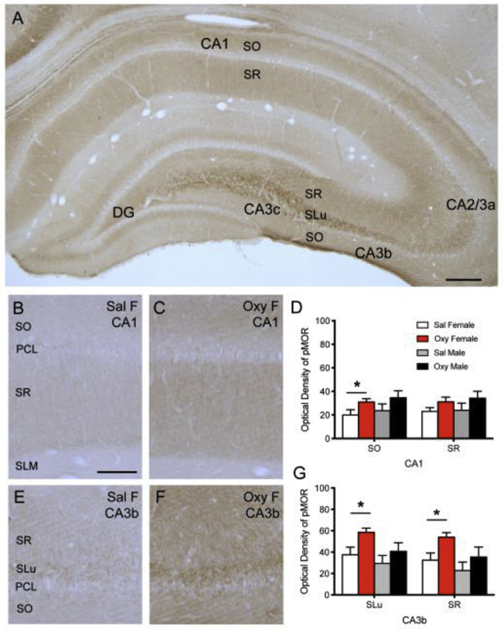
Fig 1: pMOR levels are increased in naïve Oxy-females compared to Sal-females in CA1 and CA3b.

A. pMOR-ir is most dense in the SLu of CA3a, b and c and less dense in the SO and SR of the CA1 and CA3. Sparse diffuse pMOR-ir is seen in the central hilus of the dentate gyrus (DG). B,C. Representative images show pMOR-ir in the SO, PCL, SR, and SLM of CA1 in a Sal-female (B) and an Oxy-female (C) rat. D. The density of pMOR-ir increases in the SO of CA1 in Oxy-females compared to Sal-females. E,F. Representative images of pMOR-ir in the SR, SLu, PCL, and SO of CA3b in a Sal-female (E) and an Oxy-female (F) rat. G. The density of pMOR-ir increases in the SLu and SR of CA3b in Oxy-females compared to Sal-females. Scale bar A = 250 μm; B,C,E,F = 100 μm; *p < 0.05.
