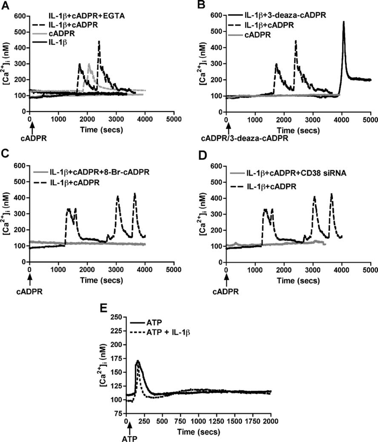
Fig. 4.

Effect of pharmacological agents and CD38 RNAi on Ca2+ dysregulation in human astrocytes. Primary astrocytes were treated with or without IL-1β and various cADPR-related pharmacological agents were applied. Single-cell Ca2+ transients were measured on Fura 2AM loaded with or without IL-1β activated astrocytes after application of various cADPR-related pharmacological agents, and the representative traces are shown. Subpanel A shows representative traces of [Ca2+]i levels in control and IL-1β-activated astrocytes after stimulation with cADPR (125 μM). We found increased basal [Ca2+]i levels upon IL-1β-activation that increased significantly by cADPR in the presence or absence of Ca2+ chelator EGTA (5 mM). Subpanel B shows a more potent increase in [Ca2+]i levels in activated astrocytes upon 10 μM 3-deaza-cADPR (non-hydrolyzable analog of cADPR) treatment over cADPR itself. In addition, both 8-Br-cADPR (100 μM), a specific cADPR antagonist (C), and CD38 siRNA (D) significantly downregulated the cADPR-mediated increase in [Ca2+]i levels. This indicates the response to be CD38-cADPR specific. Subpanel E shows that ATP-mediated Ca2+ flux in control and IL-1β-activation astrocytes were comparable
