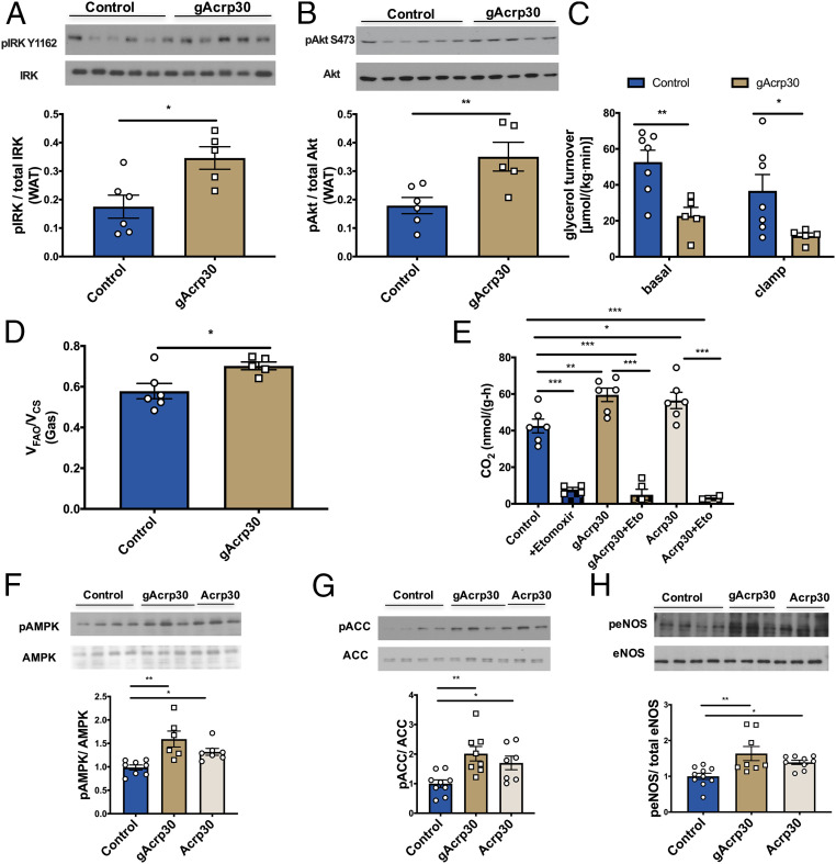
Fig. 3.
gAcrp30 improves insulin signaling in WAT and increases the switch from glucose to fat oxidation in skeletal muscle in vivo. (A) Western blot images for insulin receptor kinase phosphorylation (pY1162) in WAT (n = 5–6) in the clamp state. Quantification is shown below. (B) Western blot images for Akt phosphorylation (pS473) in WAT (n = 5–6) in the clamp state. Quantification is shown below. (C) Glycerol turnover rate under basal and hyperinsulinemic-euglycemic conditions (n = 5–7). (D) Ratio of mitochondrial ketone oxidation and β-oxidation (VFAO) to citrate synthase flux (VCS) in soleus muscle (n = 5–6). (E) Fatty acid oxidation rates of solus muscles with no treatment (control), control + etomoxir, gAcrp30 treatment, gAcrp30 + etomoxir, Acrp30 treatment, Acrp30 + etomoxir (n = 2–6). (F–H) Representative Western blot images for nontreated, gAcrp30-treated, and Acrp30-treated AMPK, ACC, and endothelial nitric-oxide synthase phosphorylation in soleus muscle. Quantification is shown below. Data are shown as mean ± SEM *P < 0.05, **P < 0.01 by two-way ANOVA with Dunnett multiple comparisons for C. *P < 0.05, **P < 0.01, ***P < 0.001 by one-way ANOVA with Tukey multiple comparisons for E–H. *P < 0.05, **P < 0.01 by unpaired Student’s t test for other graphs.

