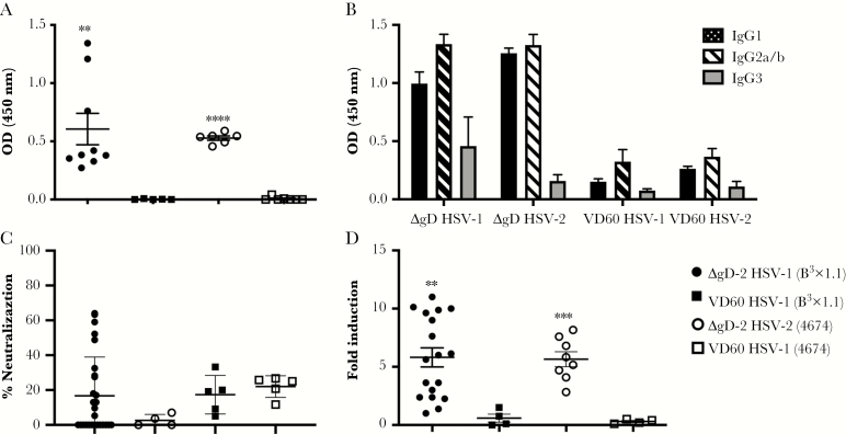
Figure 5.
Characterization of herpes simplex virus (HSV)-specific antibodies transferred from immunized dams to 7-day-old pups. Seven-day-old pups born to either ΔgD-2 or VD60 lysate-immunized dams were challenged intranasally with 105 plaque-forming units (pfu) of HSV-1(B3x1.1) or 103 pfu of HSV-2(4674). Serum samples were collected at time of sacrifice or Day 14 postchallenge and assessed as follows: (A) HSV-1- (closed symbols) or HSV-2- (open symbols) specific immunoglobulin G (IgG) was quantified by enzyme-linked immunosorbent assay (ELISA) at 1:1000 dilution with results presented as optical densitometry (OD) units. (B). The subtypes (IgG1, IgG2, or IgG3) of the HSV-specific antibodies (Abs) were quantified by a similar ELISA assay but with subtype-specific secondary Abs. Results for 1:1000 dilution of murine serum are shown as mean + standard error of the mean ([SEM] n = 4–6 mice per group). (C) The ability of the serum to neutralize HSV-1 or HSV-2 was determined by plaque reduction assay; results with 1:5 dilution of serum are shown as percentage inhibition of viral plaques relative to control (no immune serum), and each dot represents results for an individual mouse. (D) Murine FcγRIV activation was measured using a reporter bioassay with HSV-1- or HSV-2-infected targets and 1:5 dilution of serum. Each point represents results for an individual pup, and lines are mean ± SEM. Fold induction was compared by t test for immune serum from ΔgD-2- or VD60-immunized mice challenged with the indicated serotype (**, P < .01; ***, P < .001; ****, P < .0001).

