Fig. 1.
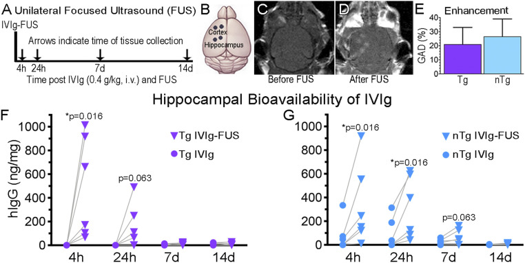
FUS increases the bioavailability of IVIg to the hippocampus. (A–D) A unilateral FUS treatment. FUS was done on the left side of the brain. (A) IVIg (0.4 g/kg) was injected intravenously in Tg and nTg animals. Animals were killed at 4 h, 24 h, 7 d, and 14 d and brain homogenates were analyzed using human IgG (hIgG) ELISA. (B) The BBB was modulated with two FUS spots (black dots) per regions, namely the cortex and hippocampus. The contralateral regions on the right side of the brain served as controls exposed to circulating IVIg without FUS permeabilization. (C and D) MRI visualization of the brain (C) before and (D) after FUS-BBB opening, which results in noticeable GAD entry as two lighter spots over the cortex, and two over the hippocampus. (E) No significant difference in GAD enhancement post-FUS, indicative of BBB permeability, was observed between Tg and nTg animals (n = 16, P = 0.21). hIgG content was found to be higher in FUS-treated hippocampi (Left, triangles) compared to the untreated hippocampi (Right, circles) in Tg mice at 4 h after IVIg-FUS delivery (F, *P = 0.016, n = 6), and in nTg mice at 4 and 24 h treatment (G, *P = 0.016, n = 6 per time-point). GAD enhancement is represented as the mean+SD of all data points per group, with no statistical difference observed between groups. Bioavailability of IVIg at each independent time-point was analyzed with a Wilcoxon matched-pairs signed rank one-tail test, under the assumption that greater levels of IVIg will be found in FUS-treated hippocampi. Significant differences were noted at P < 0.05.

