Fig. 2.
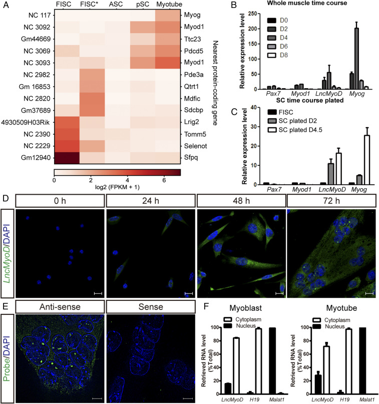
LncMyoD is an lncRNA expressed in ASCs. (A) Heat map showing the expression pattern of identified lncRNAs between RNA-seq data from different RNA-seq samples with their nearby protein-coding genes. FPKM, fragments per kilobase of transcript per million mapped reads. (B) Temporal expression of LncMyoD in whole-TA muscle extract and during muscle regeneration (D0, uninjured; D2 to D8, days 2 to 8 after muscle injury). (C) Temporal expression of LncMyoD in FACS-sorted FISCs and SCs plated in vitro under differentiation condition for 2 or 4.5 d after isolation. (D) FISH of endogenous LncMyoD molecules (green) in SCs cultured under differentiation condition for different time. The cell nuclei were stained with 4′,6-diamidino-2-phenylindole (DAPI) (blue). (Scale bar: 10 μm.) (E) FISH of LncMyoD antisense and sense probes in SCs cultured under differentiation condition for 72 h. The cell nuclei were stained with DAPI (blue). (Scale bar: 5 μm.) (F) qRT-PCR results of LncMyoD expression level in cytoplasm and nucleus in myoblasts and myotubes. H19 is used as a marker for cytoplasmic fraction; Malat1 is used as a marker for nuclear fraction.

