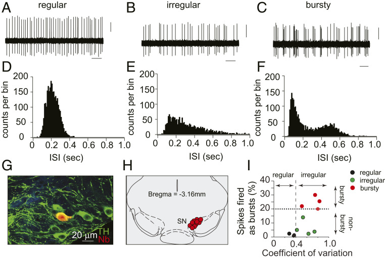
Fig. 1.
In vivo electrophysiological recordings from SN dopaminergic neurons in lightly anesthetized mice. (A–C) Representative extracellular in vivo recording of spontaneous firing activity of SN dopaminergic neurons, showing examples of regular (pacemaker, A), irregular (B), and burst firing (C) patterns. (Scale bars: 1 s and 5 mV.) (D–F) Representative histograms of interspike intrevals (ISIs) of individual regular (D), irregular (E), and bursty (F) neurons, demonstrating variations in firing patterns. (G) In vivo juxtacellular and immunocytochemical labels showing the neurochemical identity and anatomical localization of recorded dopaminergic neurons. Confocal laser scanning microscopic images of mouse brain tissue show neurons immunolabeled for TH (green) to identify dopaminergic neurons and neurobiotin (Nb, red) to visualize a neuron near the recording micropipette. (H) Anatomical mapping of all recorded WT DA neurons (n = 11) and their localization within the SN [coronal midbrain image adapted from Franklin and Paxinos’ The Mouse Brain in Stereotaxic Coordinates, Fourth Edition (58)]. (I) Scatter plot showing the coefficient of variation (mean/SD) and a fraction of spikes fired as bursts (SFB) of identified midbrain DA neurons. A dotted line at spikes fired as bursts (SFB) 20% represents the threshold for neurons classified as bursty together with respective autocorrelogram-based classifications. Note that ∼35% of the SN dopaminergic neurons exhibited burst firing under isoflurane anesthesia.

