Fig. 2.
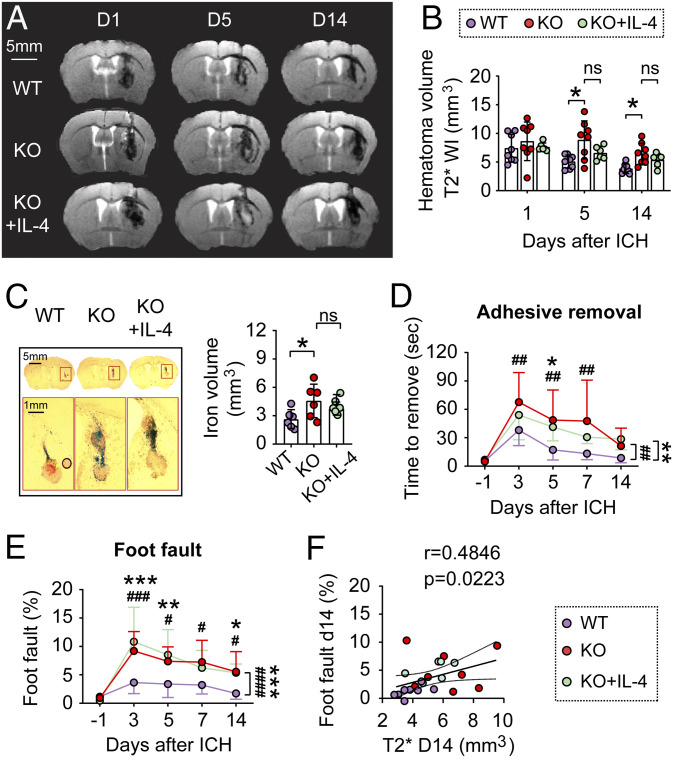
STAT6 promotes hematoma resolution and neurofunctional recovery after ICH in the blood-injection model. ICH was induced in WT and STAT6 KO mice by blood injection into the right striatum. IL-4 nanoparticles were intranasally applied starting 2 h after ICH and this was repeated daily for the next 7 d. (A) Representative T2* images obtained from the same mouse in each group at 1, 5, and 14 d after ICH. (B) Quantification of hematoma volumes on T2*-weighted images at 1, 5, and 14 d after ICH. n = 6 to 8 per group. Note that STAT6 KO mice had significantly larger hematoma volumes compared to WT mice at 5 and 14 d after ICH; meanwhile IL-4 treatment failed to significantly reduce hematoma volumes in STAT6 KO mice. (C, Left) Coronal brain sections stained for iron deposition at 14 d after ICH. (Right) Quantification of iron+-staining volume. n = 6 per group. (D and E) Sensorimotor functions were assessed by adhesive removal (D) and foot fault (E) tests before surgery (−1 d), and 3 to 14 d after ICH. n = 6 to 8 per group. (F) Spearman correlation analysis between foot fault behavior test and T2* imaging-based hematoma volumes at 14 d after ICH. ns, not significant; *P < 0.05, **P < 0.01, ***P < 0.001 KO vs. WT, #P < 0.05, ##P < 0.01, ###P < 0.001, ####P < 0.0001 KO+IL-4 vs. WT, by one-way ANOVA and Tukey post hoc tests (C) or by two-way repeated measures ANOVA (B, D, and E) and Bonferroni post hoc tests. The asterisks/pound signs at the right indicate the group differences. The asterisks/pound signs on the top indicate single day comparison between the two groups.

