Fig. 7.
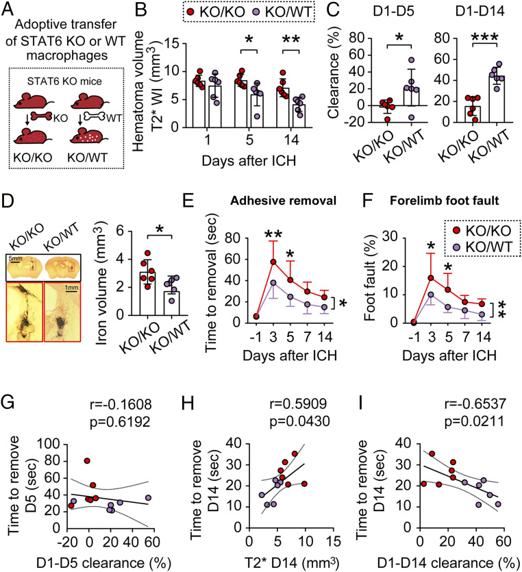
Adoptive transfer of STAT6-competent macrophages improves outcomes after ICH in STAT6 KO mice. ICH was induced in STAT6 KO mice by blood injection into the right striatum. (A) Two million macrophages prepared from either WT mice or STAT6 KO mice were adoptively transferred (intravenous route) into the STAT6 KO mice 2 h after ICH. (B) Quantification of hematoma volumes on T2*-weighted images at 1, 5, and 14 d after ICH. n = 6 per group. (C) Hematoma clearance was calculated on T2* images as the percentage decreases in hematoma size compared to the starting size on day 1. n = 6 per group. (D, Left) Coronal brain sections stained for iron deposition 14 d after ICH. (Right) Quantification of iron+ volume. n = 6 per group. (E and F) Sensorimotor functions were assessed by the adhesive removal (E) and foot fault (F) tests before surgery (−1 d) and 3 to 14 d after ICH. n = 6 per group. *P < 0.05, **P < 0.01, ***P < 0.001 by Student’s t test (C and D) or by two-way repeated measures ANOVA and Bonferroni post hoc tests (B, E, and F). The asterisks at the right indicate the group differences. The asterisks on the top indicate single day comparison between the two groups. (G) Pearson correlation analysis between the functional performance in adhesive removal test and the hematoma clearance 5 d after ICH. (H and I) Pearson correlation analysis between the functional performance in adhesive removal test and the hematoma volumes in T2*-weighted imaging 14 d after ICH (H) or the hematoma clearance 14 d after ICH (I).

