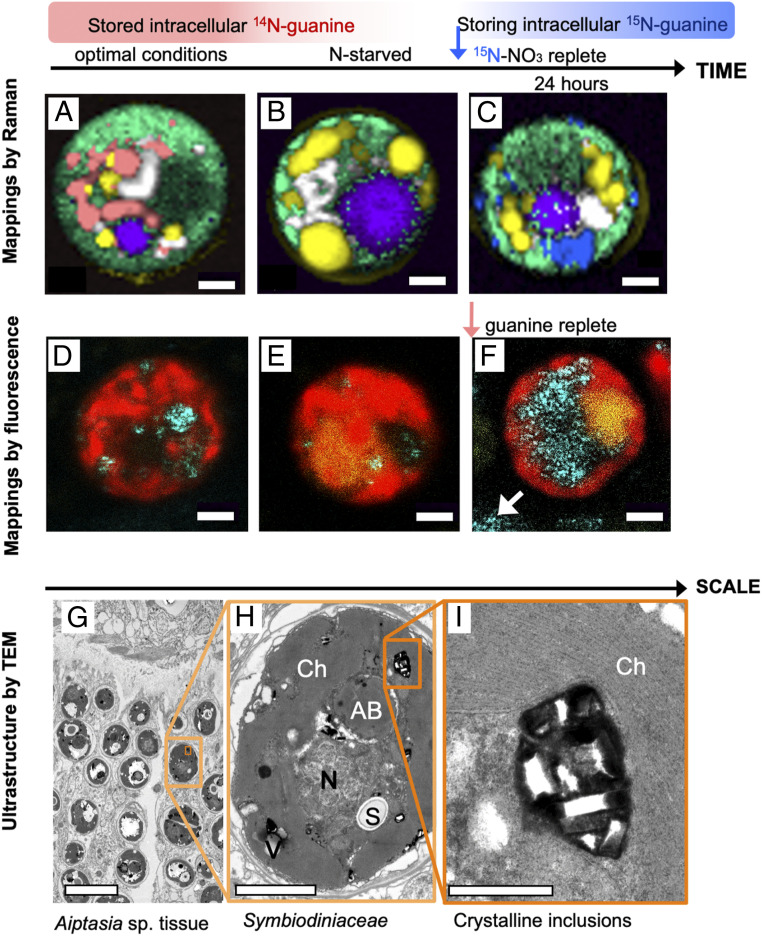
Fig. 7.
Guanine in the endosymbiotic Symbiodiniaceae cells in corals. Raman maps of cells from tissue of the coral E. paraancora (A–C). The spectra used to construct these maps are shown in SI Appendix, Fig. S13. Symbionts in corals maintained under optimal nutrient conditions contained guanine crystals (A). Cells from corals that were kept in N-depleted seawater for 4 mo contained very few guanine crystals (B). Twenty-four hours after feeding the starved coral 0.3-mM 15N-NaNO3, cells showed large 15N-guanine depots (C). The false color coding is the same as that in Figs. 1 and 4 with magenta added to represent accumulation bodies. (Scale bar, 2 μm.) Symbiodiniaceae cells isolated from the Great Barrier Reef (GBR) coral, A. millepora (D–F). Cell from freshly collected coral (D), N-depleted coral (E), and from one day after feeding with medium containing traces of undissolved guanine grains (F). Cyan, 488-nm laser reflection of guanine grains outside (white arrow) and inside the cells; red, chlorophyll autofluorescence at 670–700 nm; yellow, fluorescence in accumulation bodies at 500–560 nm; white arrow, remains of undissolved crystalline guanine in medium. (Scale bar, 2 μm.) Ultrastructure of Symbiodiniaceae cells in Aiptasia sp. shown at increasing magnification (G–I). AB, accumulation body; Ch, chloroplast; N, nucleus; S, floridean starch; V, vacuole. (Scale bars, 10 [G], 2 [H], and 0.5 μm [I].)

