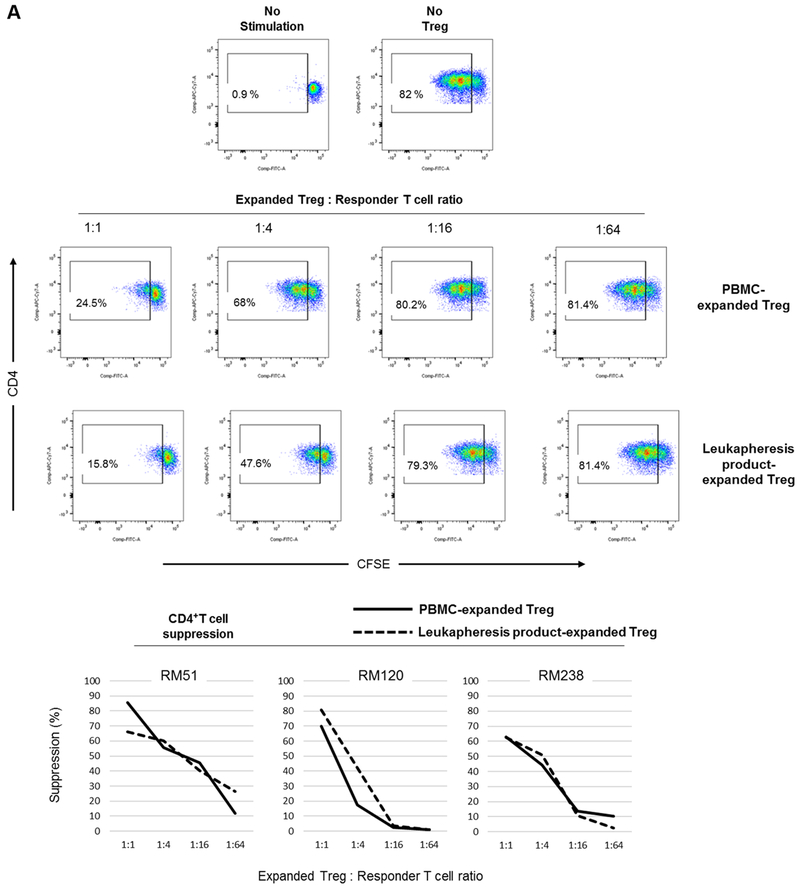
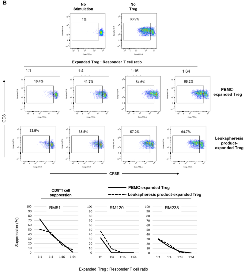
FIGURE 5. Suppressive function of Treg expanded from normal steady state PBMC before and in leukapheresis products after GM-CSF/G-CSF administration.
Treg were evaluated for their suppressive effect on autologous CD4+ (A) and CD8+ (B) T cell proliferative responses following polyclonal stimulation (n=3 monkeys). Autologous CFSE-labeled responder CD2+ T cells were stimulated by αCD2/CD3/CD28-coated microbeads at a cell:bead ratio of 1:2 for three days in the presence or absence of VPD450-labeled Treg at the indicated ratios. Percent T cell proliferation was determined by CFSE-dilution. Dot plots (upper panels) are from one animal representative of three monkeys. Graphs (lower panels) depict data from all three animals.


