
Fig. 4. Engraftment of functional hepatic hydrogel carriers.
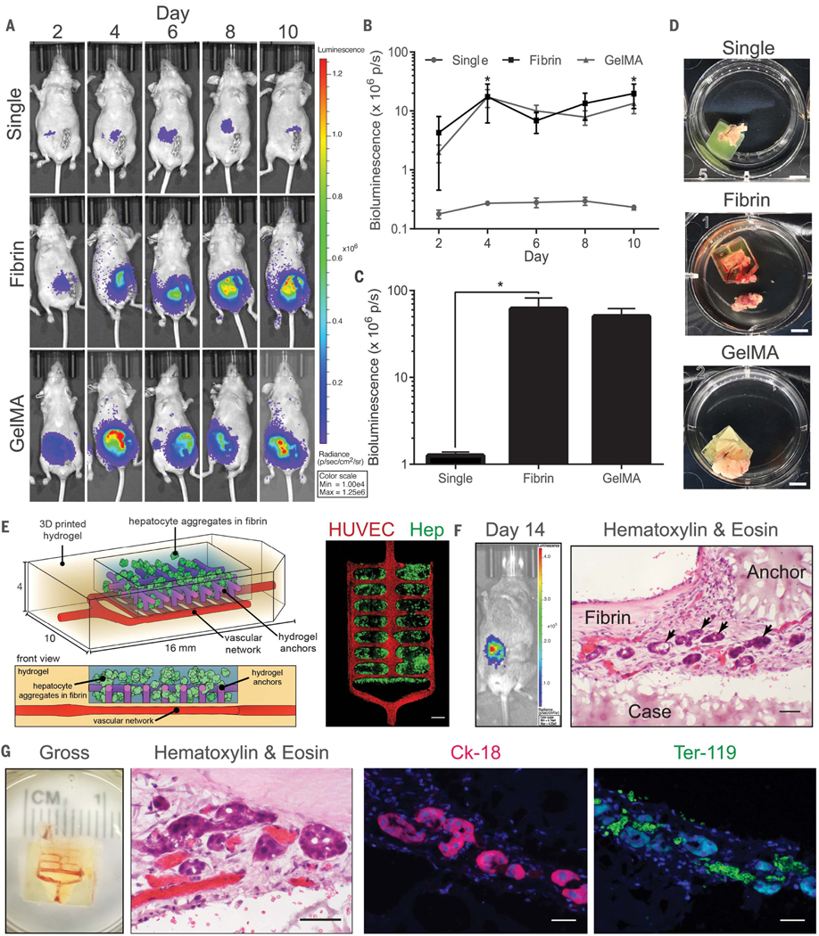
(A to C) Albumin promoter activity was enhanced in hydrogel carriers containing hepatic aggregates after implantation in nude mice. Data from all time points for each condition are shown in (B) [N = 4, *P < 0.05 by two-way analysis of variance (ANOVA) followed by Tukey’s post-hoc test]. Cumulative bioluminescence for each condition is shown in (C) (N = 4, *P < 0.05 by one-way ANOVA followed by Tukey’s post-hoc test). Error bars indicate SEM. GelMA, gelatin methacrylate. (D) Gross images of hydrogels upon resection (scale bars, 5 mm). (E) (Left) Prevascularized hepatic hydrogel carriers are created by seeding endothelial cells (HUVECs) in the vascular network after printing. (Right) Confocal microscopy observations show that hydrogel anchors physically entrap fibrin gel containing the hepatocyte aggregates (Hep) (scale bar, 1 mm). (F) Hepatocytes in prevascularized hepatic hydrogel carriers exhibit albumin promoter activity after implantation in mice with chronic liver injury. Graft sections stained with H&E show positioning of hepatic aggregates (black arrows) relative to printed (case, anchor) and nonprinted (fibrin) components of the carrier system (scale bar, 50 μm). (G) Hydrogel carriers are infiltrated with host blood (gross, H&E). Carriers contain aggregates that express the marker cytokeratin-18 (Ck-18) and are in close proximity to Ter-119–positive RBCs (scale bars, 40 μm).