
Figure 2:
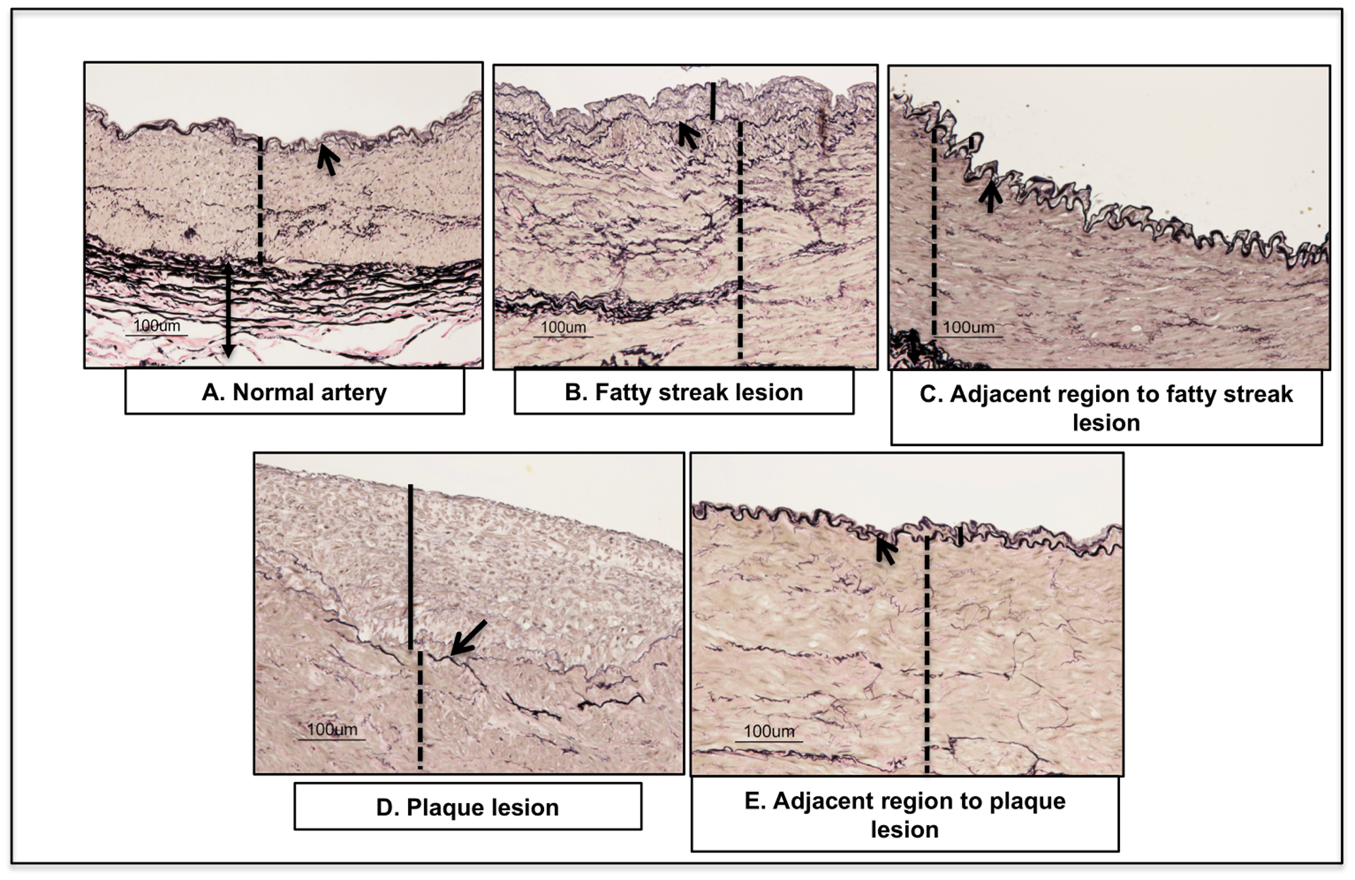
Validation of RCIA phenotypes: Cross-sections of the control, affected and adjacent regions stained with Verhaeff van Gieson displaying arterial layers: intima, and media layers). Arrow indicates fiber-like internal and external elastin lamina; arrow heads denote intimal. Solid line indicates the intimal layer; Dotted line indicates media layer.