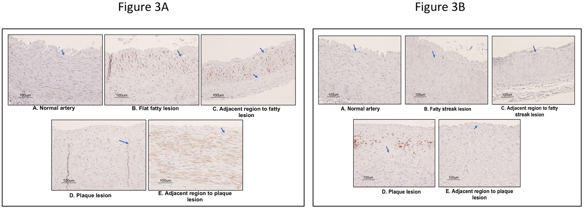
Figure 3:

(A) Detection of vascular smooth muscle cell contractile phenotype marker (SMαA) and (B) macrophage marker (CD68) in early stage atherosclerotic lesions in baboons by immunohistochemistry. Brown stain indicates the detection of SMαA or CD68. Blue stain deficit the nucleus of vascular cells. The arrow indicates the elastic lamina.
