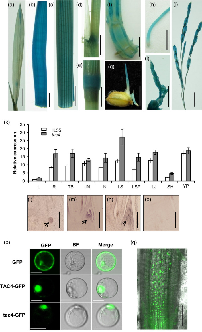
Figure 3.

TAC4 expression pattern and its product’s subcellular localization. (a–j) GUS activity levels in various organs. Leaf, (a); internode, (b); leaf sheath (c); leaf sheath pulvinus, (d); node, (e); tiller base, (f); coleoptile, (g); root, (h); young panicle (2 cm), (i); and young panicle (5–10 cm), (j). Scale bars, 1 cm. (k) The relative expression levels of TAC4 in various organs. L, leaf; R, root; TB, tiller base; IN, internode; N, node; LS, leaf sheath; LSP, leaf sheath pulvinus; LJ, lamina joint; SH, spikelet hull and YP, young panicle. (l–o) RNA in situ hybridization. Expression patterns of TAC4 were measured in the tiller bases at 30‐day after sowing. Black arrowheads indicate the positions of the tiller primordium (l) and axillary bud (m and n) in the tiller base. The sense probe was hybridized and used as the negative control (o). Scale bars, 200 μm. (p–q) Subcellular localizations of TAC4 and tac4. The TAC4‐GFP and tac4‐GFP fusion proteins are present within the nuclei in rice protoplasts (p) and in the roots of p35S:TAC4–GFP transgenic plant (q). Scale bar, 20 μm (p); Scale bar, 100 μm (q).
