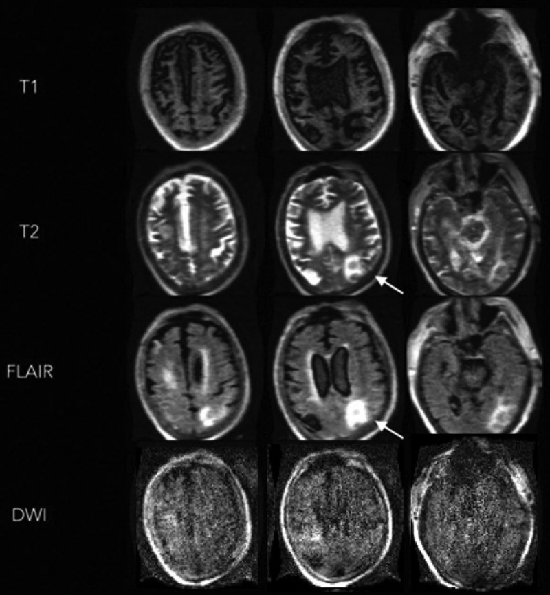
Figure 2.

Portable MRI of a critically ill patient with coronavirus disease 2019 (patient illustration 1). A 74-year-old male with a past medical history of obesity, hypertension, and diabetes was admitted with respiratory failure and was subsequently found to have severe acute respiratory syndrome coronavirus 2. The patient had a prolonged ICU course complicated by acute respiratory distress syndrome, acute kidney injury, and upper gastrointestinal bleed. A noncontrast head CT was performed after the patient failed to make a meaningful neurologic recovery despite being stabilized systemically. This revealed multifocal edema in bilateral occipital and parietal lobes, and a right superior cerebellar hemorrhage without evidence of hydrocephalus. Portable MRI-revealed diffuse atrophic changes are seen on the T1 weighted images. Hyperintense T2 weighted and fluid attenuated inversion recovery (FLAIR) signals are noted in the left parietooccipital region, without increased diffusion-weighted imaging (DWI) signal.
