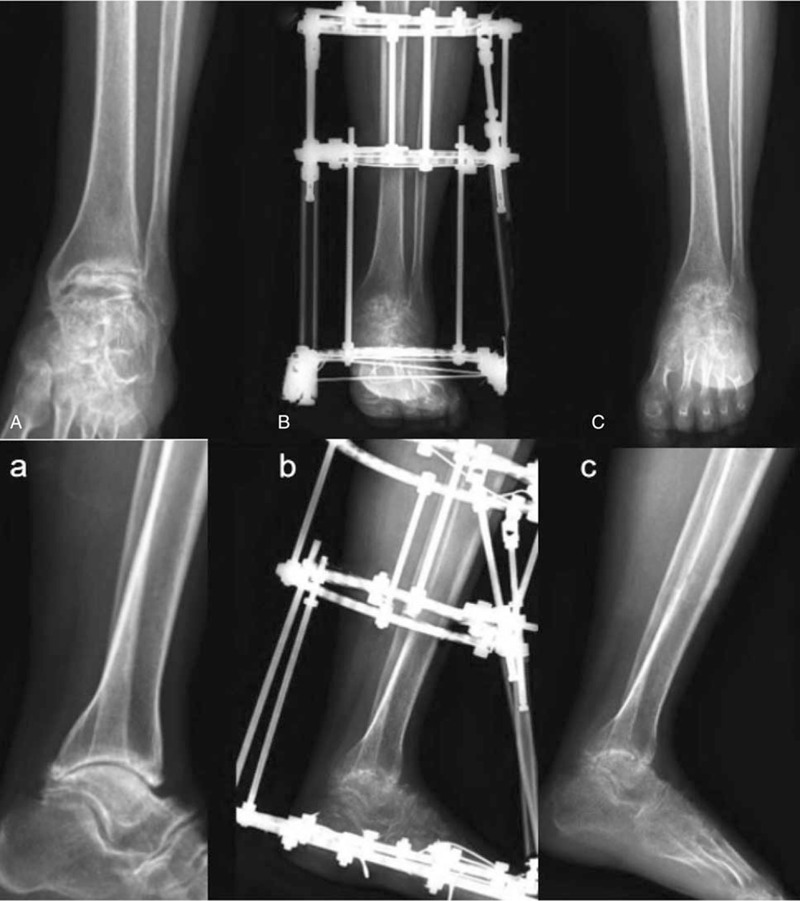
Figure 1.

(A–C) Anteroposterior X-ray of ankle joint: preoperation, postoperation, and remove the external fixator. (a–c) Lateral position: Preoperation, postoperation, and remove the external fixator. Preoperation, the joint space became narrower, the osteochondral was destroyed, osteophyte formation, and marginal hyperplasia were seen. Postoperation, the ankle joint was fused and the angle between tibiotalar joints was appropriate, and the deformity was corrected.
