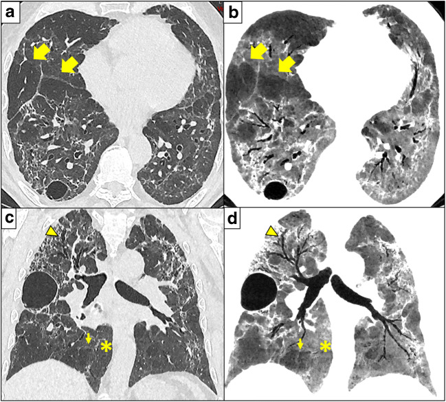
Fig. 2.
Three months after COVID-19 and ARDS, CT shows severe architectural distortion and traction bronchiectasis (arrowhead in c and d). These findings are frequent in patients that recovered from ARDS. However, multi-lobular hypoattenuation pattern is also present (arrows) on axial 1-mm-thick slices reconstructed with lung kernel (a) and 10-mm-thick minimum intensity projections (mIP) slices with soft kernel (b), and on coronal reformats 1-mm-thick reconstructed with lung kernel (c) and 10-mm-thick minimum intensity projections with soft kernel (d). The mIP post-processing (b, d) highlights a geographic, lobular hypoattenuation pattern that most likely represents multifocal small airways disease. Presumably hyperinflated lobules further exhibit convex perilobular septae towards normal lung parenchyma (small arrows) that should correspond to an air trapping on expiration. Note that the normal lung parenchyma (stars) is more easily assessed on mIP reformats with narrow window settings (b, d), than on classical lung analysis (a, c). Note pneumatoceles in subpleural location described in COVID-19 pneumonia

