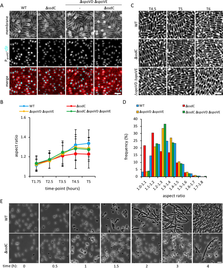
Fig 7. Forespore shape and germination in cortex mutants.
(A) Forespore morphology of wild-type (WT, bBK15), ΔssdC (bBK18), ΔspoVD ΔspoVE (bJL3) and ΔssdC ΔspoVD ΔspoVE (bJL82) strains at 4.5 h after onset of sporulation (T4.5). Forespore cytoplasm was visualised using a forespore reporter (PspoIIQ-cfp, false-coloured cyan in merged images). Cell membranes were visualised with TMA-DPH fluorescent membrane dye and are false-coloured red in merged images. Scale bar = 2 μm. (B) Average forespore aspect ratio (± STDEVP) of wild-type (WT, bBK15, blue), ΔssdC (bBK18, red), ΔspoVD ΔspoVE (bJL3, yellow) and ΔssdC ΔspoVD ΔspoVE (bJL82, green) strains during a sporulation time-course. n > 200 per time-point, per strain. (C) Phase-contrast micrographs of wild-type (WT, bBK15), ΔssdC (bBK18) and ΔspoVD ΔspoVE (bJL3) strains at T4.5, T5 and T6 of sporulation. Forespore cortex is visualised as phase-bright areas within the spore. Phase-dark spores in the ΔspoVD ΔspoVE (bJL3) mutant indicate absence of cortex. Scale bar = 2 μm. (D) Frequency distribution histogram of forespore aspect ratio of wild-type (WT, bBK15), ΔssdC (bBK18), ΔspoVD ΔspoVE (bJL3) and ΔssdC ΔspoVD ΔspoVE (bJL82) strains at 5 h after the onset of sporulation (T5), n >1000. (E) Phase-contrast micrographs of wild-type (bAT87, WT) and ΔssdC (bJL56) spores during a germination and outgrowth time-course in nutrient-rich media (LB). Scale bar = 5 μm.

