
Figure 1.
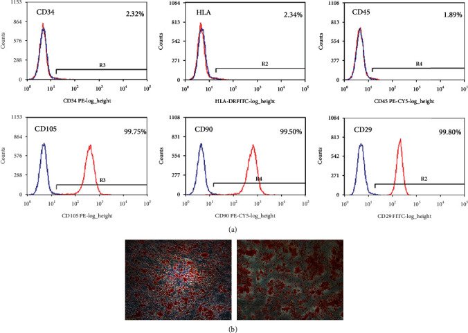
Morphological observation and characterization of ADSCs cultured in vitro. (a) Flow cytometry labeling of surface markers showed positive expression of CD29, CD90, and CD105 with positive rates of 99.98%, 99.5%, and 99.75%, respectively. CD34, CD45, and human leukocyte antigen-DR expression was negative with expression rates of 2.32%, 1.89%, and 2.34%, respectively. (b) After adipogenic induction, P3 cells were stained with Oil Red O, and oil droplet-like adipose tissue was observed by microscopy. After osteogenic induction, Alizarin Red staining showed that aggregated bone tissue was stained red in microscopy analysis.