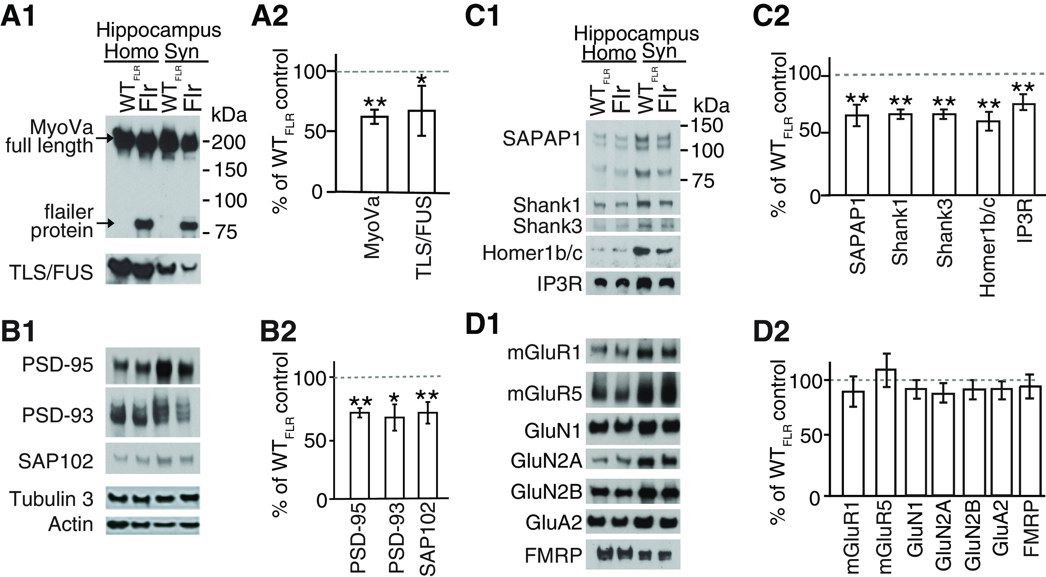
Figure 5.

Synaptic transport of proteins to PSD is altered in Flr mice. WTFLR and Flr hippocampal synaptosome extracts were used for quantitative Western blot analyses of synaptic proteins. A1, A2, Levels of full-length MyoVa and TLS/FUS were reduced in Flr extracts. B1, B2, Expression levels of major MAGUKs (PSD-95, PSD-93, SAP102) and (C1, C2) SAPAP1, SHANK1, SHANK3, Homer1b/c, and IP3R were significantly reduced in Flr. D1, D2, mGluRs, NMDAR, AMPAR subunit levels, and FMRP were not changed in Flr when compared with WTFLR. Homogenate (Homo) and synaptosome (Syn) was prepared from whole hippocampus. Protein levels in Flr were quantified and plotted as % of WTFLR; n = 3 samples per group/each sample pool three to five animals. Unpaired t test was used for statistical analysis. Error bars represent ±SEM; *p ≤ 0.05, **p ≤ 0.001.
