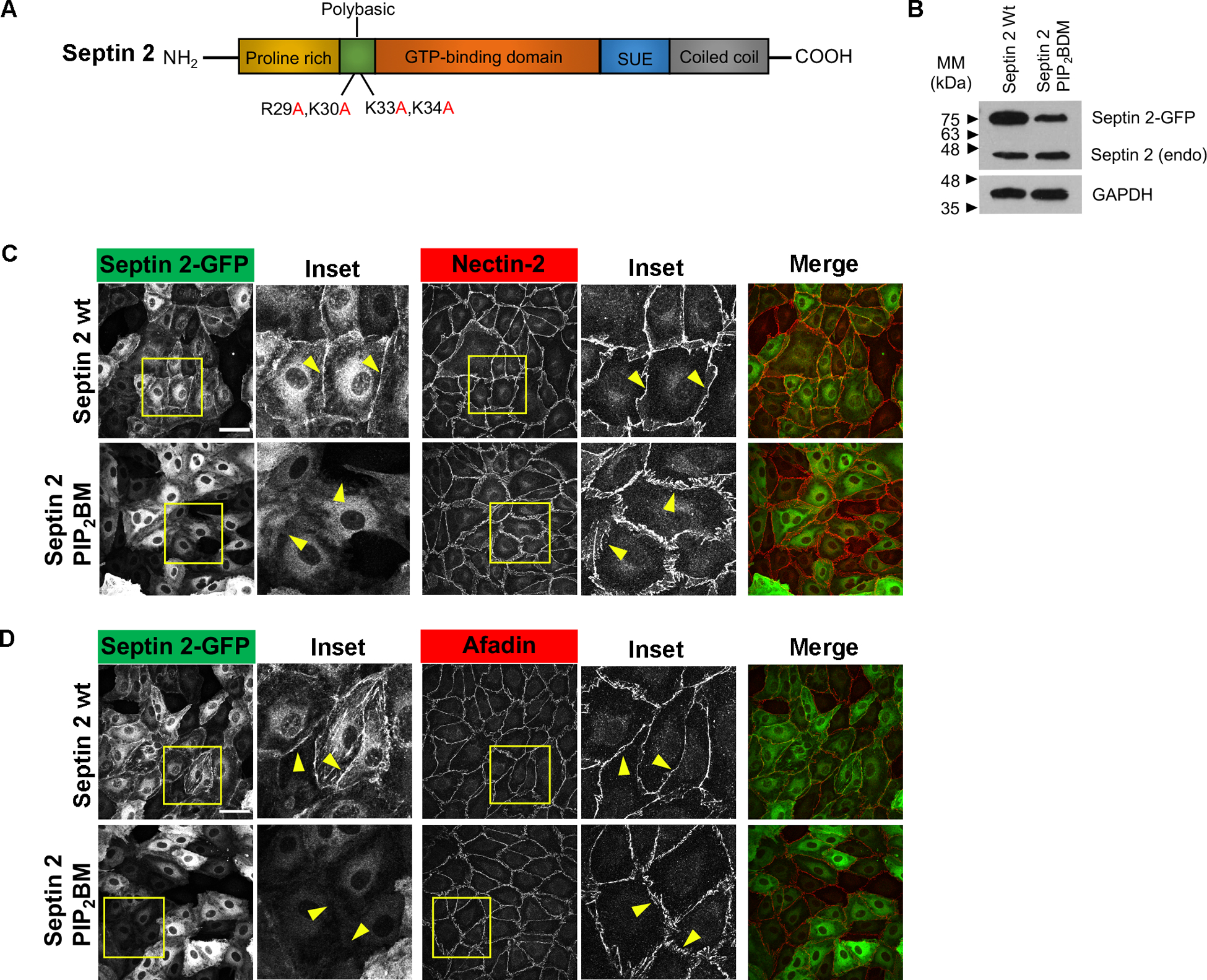
Figure 5. Polybasic mutant septin 2 fails to localize to cell junctions.

(A) Diagram of septin 2 domain architecture shows four basic residues, R and K, in the polybasic domain at the N-terminus. (B) Ectopic overexpression levels of septin 2 wild type (wt)-GFP and septin 2 PIP2 binding mutant (BM)-GFP. (C-D) Immunofluorescence staining shows overexpression of septin 2 wt and PIP2 BM (green), junctional proteins (red), and actin filaments. (C) Overexpression of septin 2 wt-GFP and septin 2 PIP2BM-GFP (Upper, Inset) and organization of nectin-2 at cell junctions (Lower, Inset). (D) Overexpression of septin 2 wt-GFP and septin 2 PIP2BM-GFP (Upper, Inset) and organization of afadin at cell junctions (Lower, Inset). Scale bars: 50 μm
