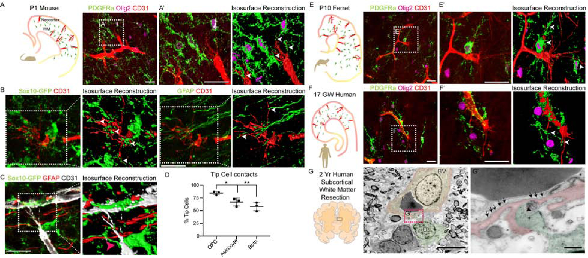
Figure 1: Conserved OPCs-Endothelial Tip Cell Physical Interactions in Developing White Matter Tracts.

(A-A’) OPC-Endothelial tip cell interactions in P1 mouse forebrain coronal section (A). OPCs (PDGFRa+Olig2+ cells) in WM frequently contact endothelial tip cell (CD31+) filopodia (A’). Isosurface reconstruction shows OPCs enwrap tip cell filopodia (white arrowheads). Scale bars = 25μm.
(B, C) Representative images and isosurface reconstructions from a P1 Sox10-GFP mouse showing CD31+ tip cell filopodia are frequently enwrapped by GFP+ OPC processes (white arrowheads) but not astroglial (GFAP+) processes. Scale bar = 20μm (D) Quantification of tip cells contacted by OPCs and astroglial cells or both in the P1 WM.
(E–E’) P10 ferret brain coronal section and isosurface reconstruction showing OPCs in WM frequently enwrap endothelial tip cell filopodia. Scale bars = 25μm. (F–F’) OPC-tip cell interactions in 17 gestational weeks (GW) human brain coronal section, showing similar findings to mouse and ferret. Scale bars = 25μm.
(G–G’) Persistent oligodendroglial-tip cell interactions shown by ultrastructural analysis of subcortical WM resection from 2-year-old human, combined with Olig2 immunogold labelling. Note oligodendroglial cell process (puesdocolor-green; black arrowhead) contacting filopodial extensions from a nascent vessel (puesdocolor-red) with caveolae (black arrows). Scale bars = 5μm (G), 500nm (G’). BV, Blood Vessel; RBC, Red Blood Cell; O, Oligodendroglia. Data are represented as mean ± S.D. and analyzed by two-tailed unpaired Student’s t test. n=3 animals. Significance: *p<0.05, **p<0.01.
