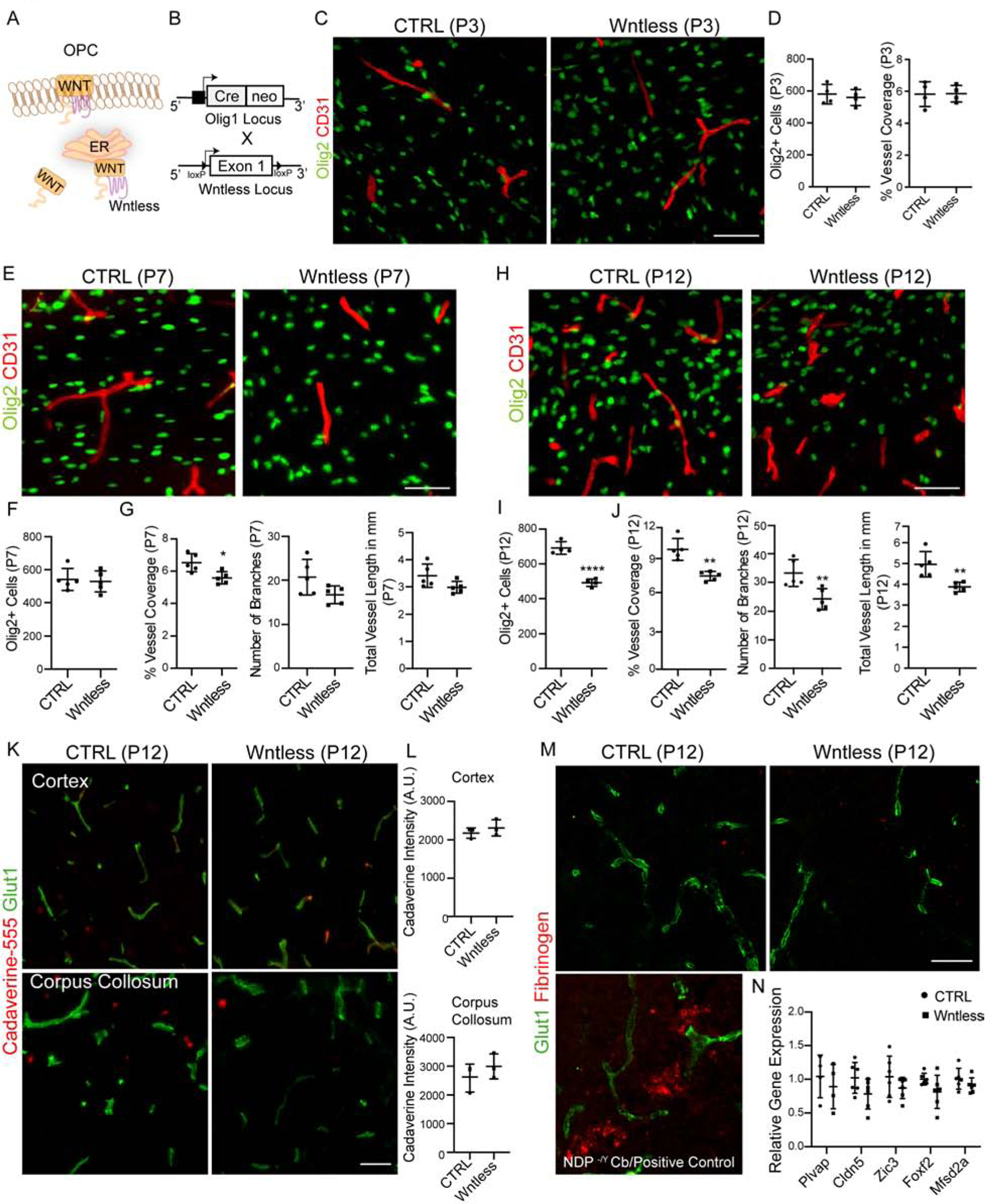
Figure 5: OPC Wnt Activity is Necessary for Postnatal White Matter Vascular Development.

(A) Wntless in the endoplasmic reticulum promotes Wnt ligand production and transport to the plasma membrane where they are secreted.
(B) Schematic of Wntless conditional knockout (Wntless cKO) generation.
(C, D) White matter BV development in Wntless cKO is not affected at P3, as shown by Olig2+ and CD31+ labeling and quantification. Scale bar = 50μm.
(E–G) Representative images from P7 cortical WM region showing Olig2+ oligodendroglial cells and CD31+ BVs and quantification. Note a decrease in BV density compared to control. Scale bar = 50μm.
(H–J) Representative images and quantification from P12 cortical WM showing significantly reduced Olig2+ oligodendroglial cells and CD31+ BV coverage in Wntless cKO. Scale bar = 50μm.
(K, L) Representative images and quantification of fluorescence intensity from cortex (top panels) and corpus collosum (bottom panels) regions of Cadaverine-555 injected mice reveal no tracer leakage indicating no BBB disruptions in Wntless cKO.
(M) Representative images of corpus collosum region from control and Wntless cKO mice labelled with Glut1 and Fibrinogen. Note absence of fibrinogen deposits in Wntless cKO mice indicating no BBB damage. Bottom left panel shows positive control for fibrinogen staining from cerebellum of Norrin (NDP −/Y) mutants.
(N) qPCR gene expression analysis of WM isolates from CTRL and Wntless cKO mice reveals no alterations in expression of genes involved in BBB maturation and maintenance. Data are represented as mean ± S.D. and analyzed by two-tailed unpaired Student’s t test. n=4 animals/genotype (D), n=6 (F–G), n=5 (I–J), n=3 (L), n=6 (N). Significance: *p<0.05, ***p<0.001, **** p<0.0001.
