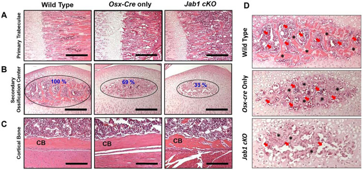
Figure 2. Jab1 cKO mice exhibited severely altered bone morphology and delayed secondary ossification center formation.
Hematoxylin and Eosin staining in the proximal tibiae of wild type, Osx-cre only, and Jab1 cKO mice at 18 days of age. (A) Representative images of primary trabeculae. Scale bars, 100μm. (B) Representative images of the secondary ossification centers (SOC). Scale bars, 100μm. (C) Representative images of the cortical bone (CB). Scale bars, 50μ.

