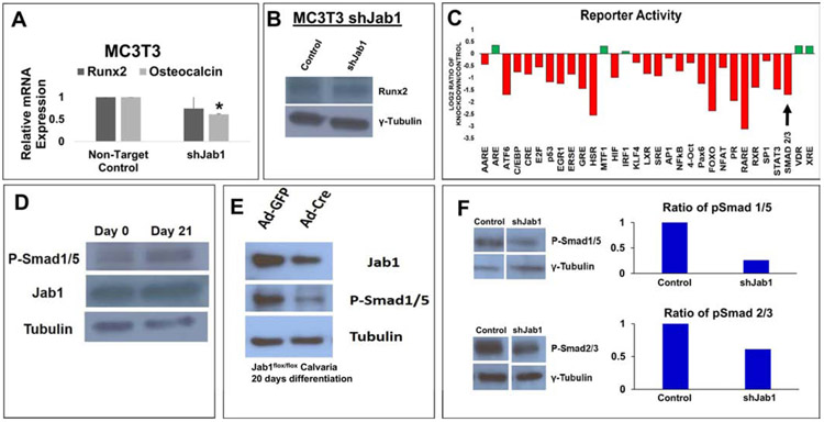
Figure 7. Jab1-knockdown inhibited osteoblast differentiation likely in part through the TGFβ/BMP signaling pathways.
(A) RNA expression levels of Runx2 and Osteocalcin in Jab1-knockdown MC3T3-E1 cells. (B) Protein expression levels of Runx2 and Jab1 in Jab1-knockdown MC3T3-E1 cells. (C) An unbiased reporter screen in Jab1-knockdown MC3T3-E1 cells. The black arrow indicates a canonical Smad2/3 TGFβ reporter. A detailed list of the pathways in this screen can be found in Supplementary Table 3. (D) Western blot analysis of Jab1 and phospho-Smad1/5/8 in primary calvarial osteoblasts cultured in osteoblast differentiation media for 0 and 21 days, respectively. (E) Western blot analysis of Jab1 and phospho-Smad1/5/8 in control and Jab1-knockdown primary calvarial osteoblasts after 21 days of culture in osteoblast differentiation media. (F) Western blot analysis of phospho-Smad1/5/8 (top) and phospho-Smad2/3 (bottom) in control and Jab1-knockdown MC3T3-E1 cells treated with 300ng/mL BMP7 or 2ng/mL TGFβ-3 for 1 hour, respectively.

