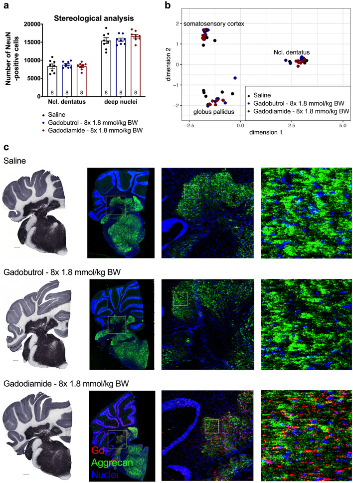
Figure 3.
No alterations in number of NeuN-positive cells and gene expression as well as overlap of aggrecan and Gd distribution in the lateral cerebellar nucleus. (a) Numbers of NeuN-positive cells in the lateral cerebellar nucleus and adjacent deep cerebellar nuclei of treated and control animals 11 weeks p.i. determined by stereological analysis. (b) Gene expression pathway analysis in the lateral cerebellar nucleus, globus pallidus and somatosensory cortex (control region) 11 weeks p.i. (c) Conventional immunohistochemistry of aggrecan in the cerebellum of animals sacrificed 11 weeks p.i. (left panel, scale bar: 500 µm). Distribution of Gd and aggrecan in the cerebellum of animals sacrificed 5 weeks p.i. by immunohistological LA-ICP-MS (right panel). Representative pseudo-colored overlays of Gd (red), aggrecan (green) and an iridium containing nucleic acid intercalator (blue) at different magnification. Fields of higher magnification are indicated by squares. Data represent mean ± SEM; n is indicated in the columns.

