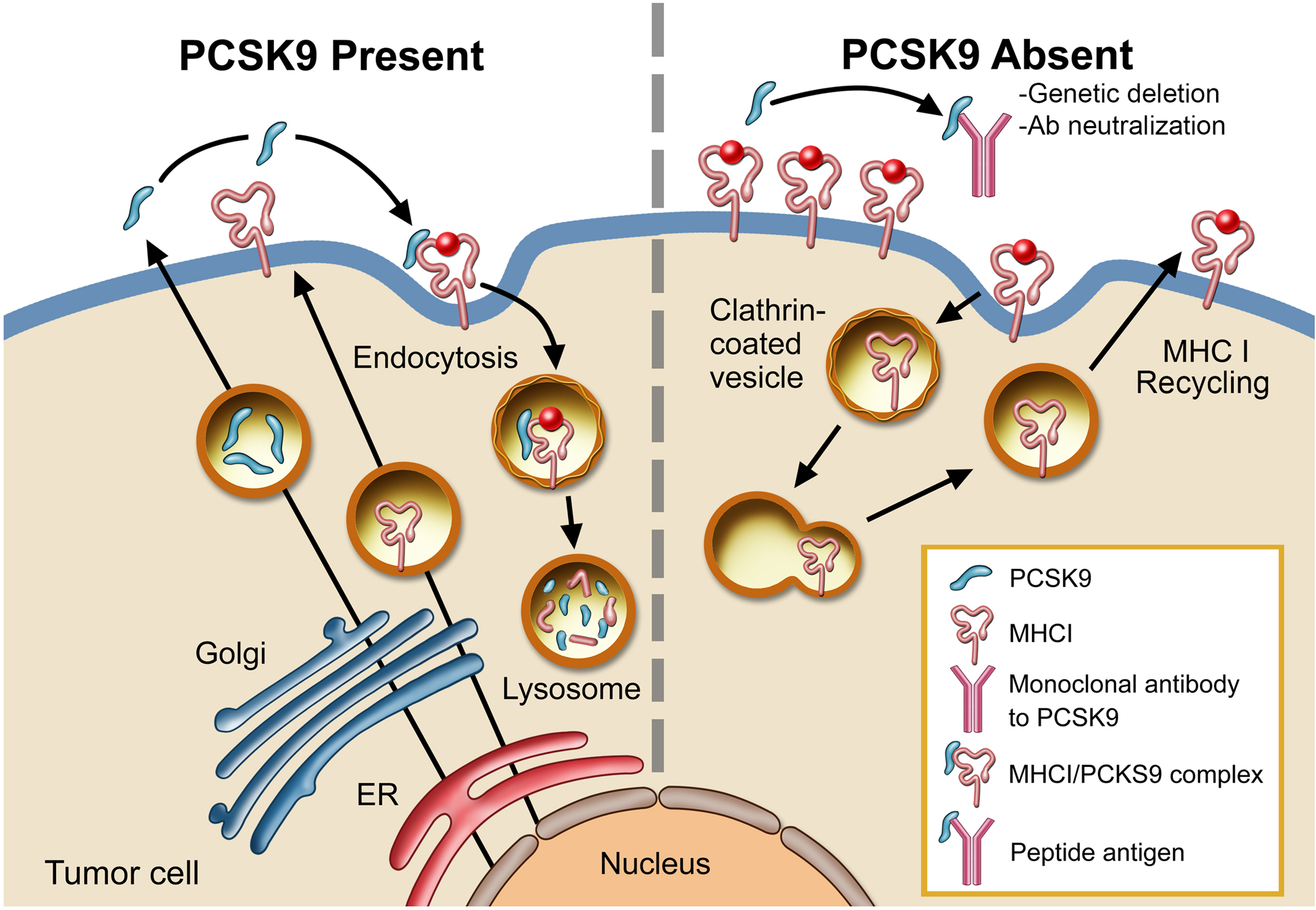
Extended Data Fig. 10. A schematic diagram illustrating PCSK9-mediated degradation of MHC I in the lysosome.

In the presence of PCSK9, MHC I is transported into the lysosome and degraded (left panel). In the absence of PCSK9, either because of genetic deletion or antibody neutralization, MHC I levels on the surface remains high and is thus able to present tumor-specific peptic antigens more efficiently to T cells (right panel). Illustration by Stan Coffman.
