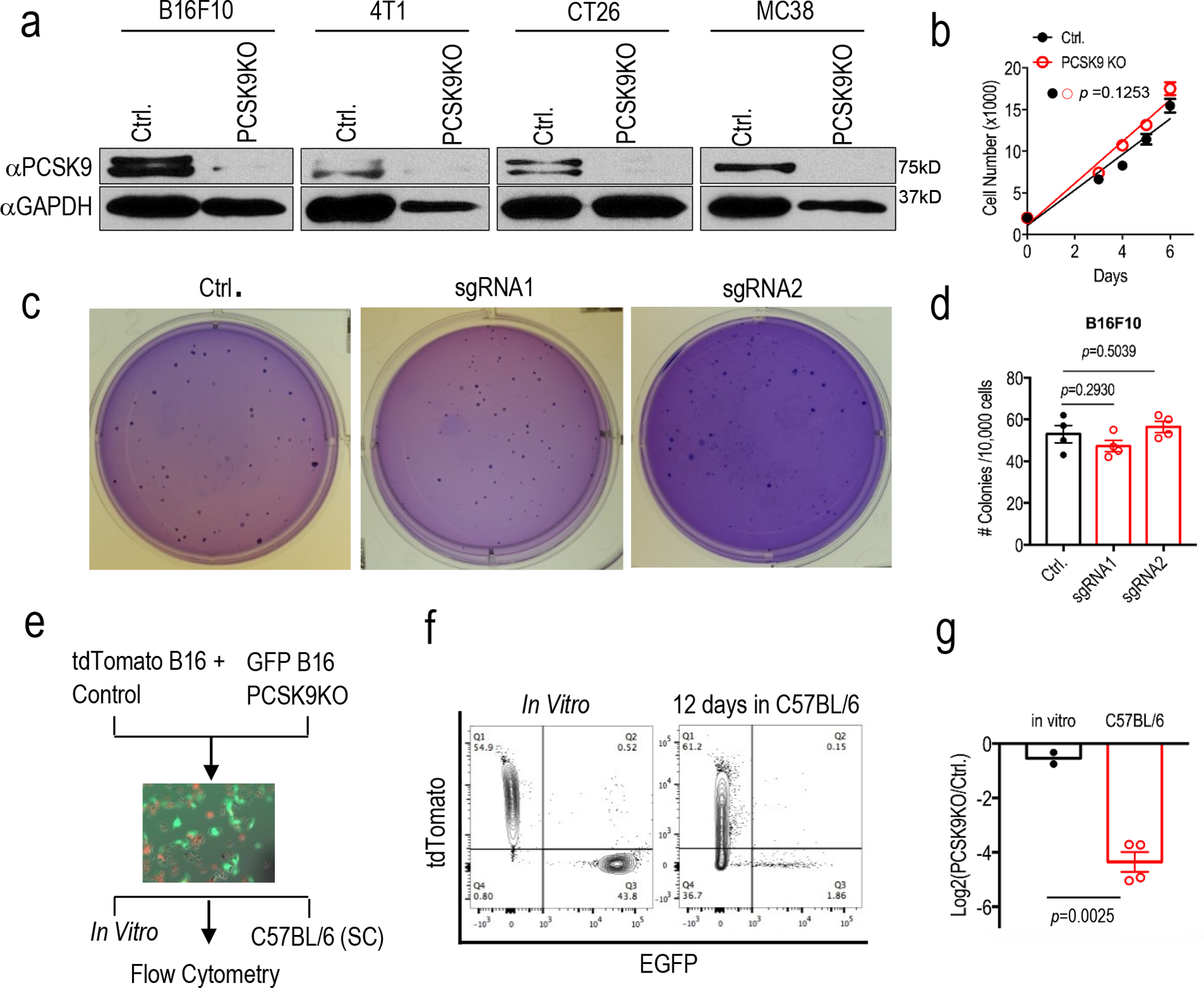
Extended Fig.1. CRISPR-Cas9 mediated knockout of PCSK9 gene and its effect on tumor cell growth in vitro and in vivo.

a. Western blot analysis of the expression of PCSK9 in murine tumor lines with PCSK9 knockout. GAPDH was used as protein loading control. Analysis was done twice with biologically independent samples. b. Cell growth of vector control or PCSK9 KO B16F10 tumor cells. Results from 5 biologically independent samples. Error bars, mean ± S.E.M. P value calculated by unpaired two-sided t test. c. Soft agar analysis of the colony formation ability of vector control or PCSK9 sgRNA-transduced B16F10 tumor cells. d. Quantitative representation of soft agar formation in c. n=4 biologically independent samples. Error bars, mean± S.E.M. P values calculated by unpaired two-sided t test. e. Diagram of in vivo competition assay. f. Change in ratios of mixed control-tdTomato and PCSK9 KO-EGFP B16F10 cells after 12 days grown in vivo (subcutaneously) in C57BL/6 mice, as determined by flow cytometry. g. Quantitative representation of the flow analysis in f. Error bar, mean± S.E.M. n=2 and 4 biologically independent tumor samples for the in vitro and in vivo groups, respectively. P value determined by unpaired two-sided t test.
