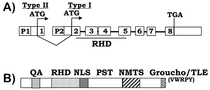
Figure 1. RUNX2 gene and protein structure.
A) RUNX2 gene structure. Two major RUNX2 isoforms are transcribed from the P1 and P2 promoters respectively, indicating by the ATG start codons, which are encoded by exon 1–8 (type II) or exons 2–8 (type I). The DNA binding Runt homology domain (RHD) is encoded by exons 2–5. B) RUNX2 protein structure. In addition to the RHD domain, RUNX2 proteins contain a glutamine/alanine (QA) rich region, a nuclear-localization signal (NLS), a proline/serine/threonine (PST) rich region, a nuclear matrix targeting signal (NMTS), and a C-terminal VWRPY domain for TLE/Groucho interactions.

