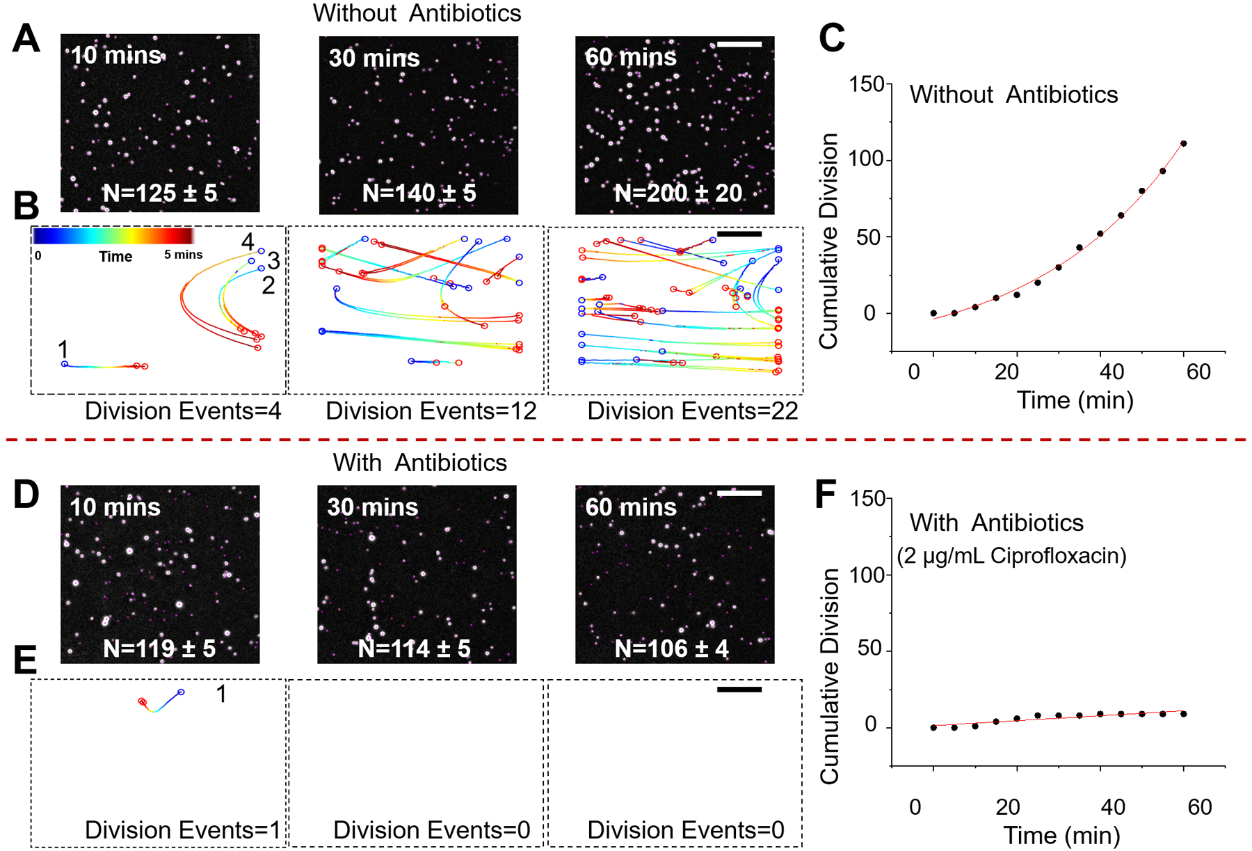
Figure 2. Validation of digital LVSi-AST with pure E. coli cultures.

Snapshots of E. coli cells after 10, 30, and 60 min in growth medium without antibiotics (A) and with antibiotics (2 μg/mL ciprofloxacin) (D). Division events over a 5-min time interval at the different time points with (B) and without ciprofloxacin (E). Cumulative division events over 60 min with (C) and without ciprofloxacin (F). Scale bar, 400 μm.
