Fig. 5.
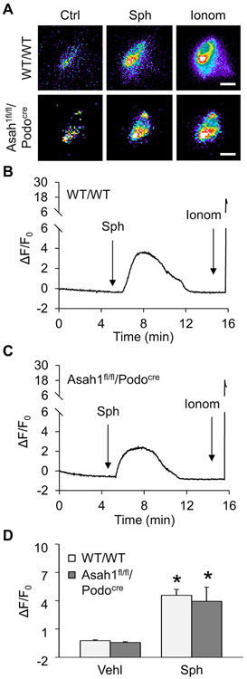
Rescue of TRPML1 channel function by Sph in podocytes with Asah1 gene deletion. A. Representative images showing that Sph (20 μM) induced TRPML1 channel-mediated Ca2+ release in podocytes of both WT/WT and Asah1fl/fl/Podocre mice. Scale bars = 40 μm. B. A representative curve showing that Sph induced elevation of GCaMP3 signal in podocytes of WT/WT mice. C. A representative curve showing that Sph induced elevation of GCaMP3 signal in podocytes of Asah1fl/fl/Podocre mice. D. Summarized data showing that Sph rescued TRPML1 channel function in podocytes lacking Asah1 gene (n = 4–5). *p < 0.05 vs. Vehl group. Ctrl, control; Sph, sphingosine; Ionom, ionomycin; Vehl, vehicle.

