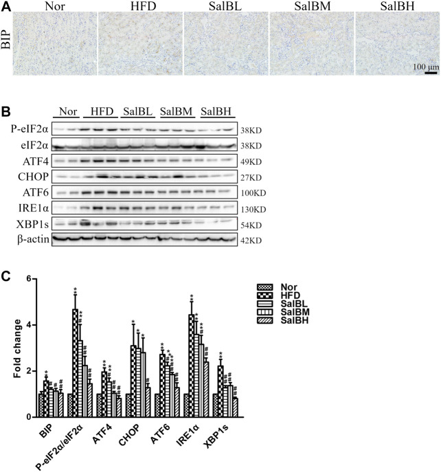
FIGURE 3.
SalB attenuated HFD-induced ER stress in the kidney cortex of mice (A,C) Representative photomicrographs of BIP-stained kidney sections and the corresponding quantitative analysis (B,C) Protein levels of P-eIF2α, eIF2α, ATF4, CHOP, ATF6, IRE1α, and XBP1s in the kidney cortex were detected by western blotting. The corresponding quantitative analysis was shown as well. Nor, mice fed with normal diet; HFD, mice fed with high-fat diet; SalBL, SalBM, or SalBH, mice fed with high-fat diet and treated with low dosage of SalB (3 mg/kg), medium dosage of SalB (6.25 mg/kg), or high dosage of SalB (12.5 mg/kg), respectively. Data are represented as means ± SEM (n = 6–8). *p < 0.05 and **p < 0.01, compared with Nor; # p < 0.05 and ## p < 0.01, compared with HFD.

