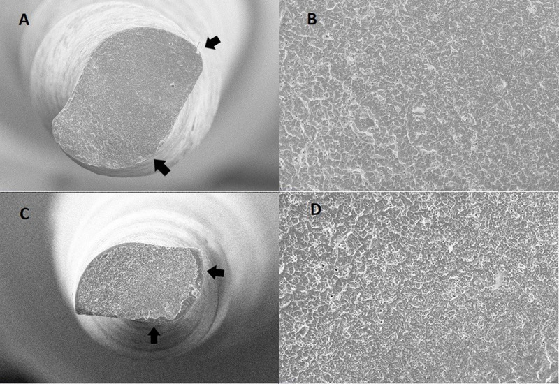
Figure 1.
Scanning electron microscopic appearances of the Reciproc Blue and VDW.rotate instruments after cyclic fatigue testing. The fracture surface view of Reciproc Blue (A), VDW.rotate (C), and a high-magnification view of the Reciproc Blue (B) and VDW.rotate (D) instruments. The crack initiation origins (arrows) are observed in the fracture surface.

