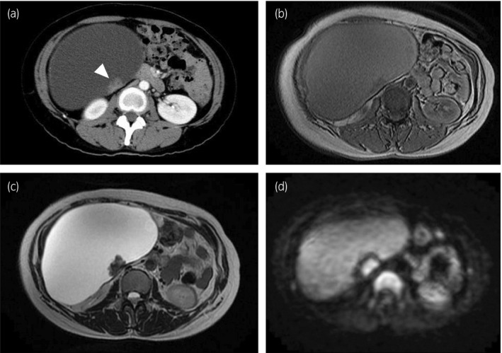
Fig. 1.

(a) Computed tomography demonstrated a large cystic tumor with enhanced solid lesion (arrowhead). Magnetic resonance imaging showed homogenous cyst on (b) T1‐weighted image and (c) T2‐weighted image. (d) Diffusion‐weighted imaging demonstrated high‐signal intensity in the solid lesion.
