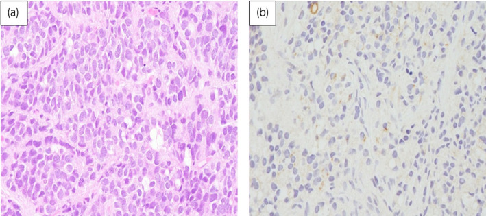
Fig. 2.

Microscopic features of the prostate biopsy specimens (hematoxylin and eosin staining). The specimens were occupied by tumor cells which had large round nuclei with prominent nucleoli and proliferated in a sheet‐like pattern (a), and weakly positive for PSA staining (b).
