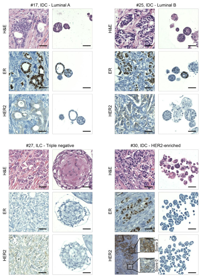
Figure 2.
Organoids recapitulate the histological features of primary BCs. Representative images of hematoxylin/eosin staining (H&E) and immunohistochemical analyses on sections of BCs and derived organoids. From left to right and from the top to the bottom are examples of luminal A, luminal B, triple negative, and HER2-enriched BCs, respectively; IDC, invasive ductal carcinoma; ILC, invasive lobular carcinoma; ER, estrogen receptor; HER2, human epidermal growth factor receptor 2. Magnifications of HER2 staining in different areas of cancer tissue #30 are also shown. Scale bar, 100 μm.

