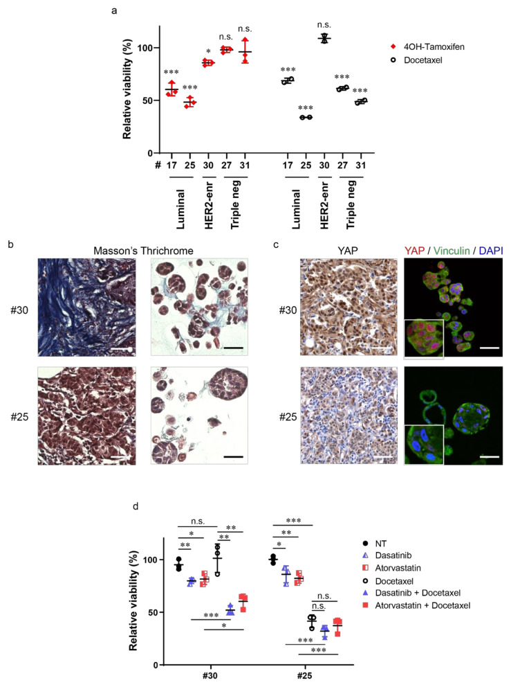
Figure 4.
Tumor organoids represent a powerful model to evaluate novel potential therapeutic strategies. (a) Dot plot showing relative viability of BC organoids treated with either 10 μM 4-Hydroxytamoxifen (4OH-Tamoxifen) or 1 nM Docetaxel for 7 days. Data are normalized to control treatment (solvent), set to 100%. Ethanol was used as control for 4-Hydroxytamoxifen, while dimethyl sulfoxide (DMSO) was used for Docetaxel. Data shown are the means ± s.d. of n = 3 (4OH-Tamoxifen) and n = 2 (Docetaxel) independent experiments; * p < 0.05, *** p < 0.001; n.s., not significant; t-test, using the Holm–Sidak method. (b) Representative images of Masson’s trichrome staining on sections of BC (left) and derived organoids (right) of the indicated cases. Scale bar, 50 μm. (c) Representative images of Yes-associated protein 1 (YAP) immunohistochemistry on BC sections (left) and of YAP (red) and Vinculin (green) immunofluorescence on sections of the derived organoids of the same cases (right). Nuclei were counterstained with either hematoxylin (left) or 4′,6-diamidino-2-phenylindole (DAPI) (right). Scale bar, 50 μm. Magnifications of YAP/Vinculin staining are also shown. (d) Dot plot showing relative viability of BC organoids treated with 100 nM Dasatinib, 100 nM Atorvastatin, 1 nM Docetaxel, their combinations, or solvent (NT, DMSO) for 7 days. Data shown are the means ± s.d. of n = 3 independent experiments, * p < 0.05, ** p < 0.01, *** p < 0.001; n.s., not significant; t-test, using the Holm–Sidak method.

