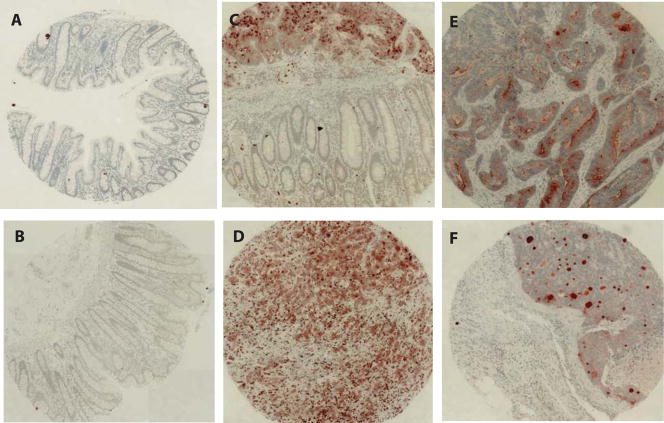
Figure 1.
NGAL expression in colon carcinoma tissues.
Tissue microarray consisting of 16 non-neoplastic colorectal mucosal specimens and 19 colorectal carcinoma tissues was constructed. The section was stained for the expression of NGAL using standard immunohistochemistry procedures. A–B: Absence of staining for NGAL in the non-neoplastic mucosa. C: A tissue section containing both of carcinoma and non-neoplastic mucosa. NGAL is evident only in the carcinoma tissue. D–F: Examples of staining for NGAL in the colorectal carcinomas.

