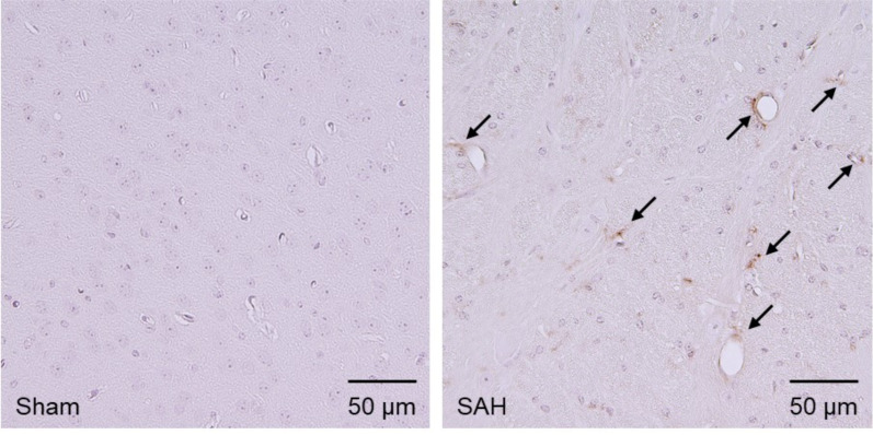
Fig. (4).
Immunohistochemical staining for immunoglobulin G (IgG) in the left temporal cortex at 1.0 mm posterior to the bregma at 24 hours after subarachnoid hemorrhage (SAH) induction in mice. Compared with sham-operated mice, extravasated IgG is stained in SAH-operated mice (arrows). (A higher resolution / colour version of this figure is available in the electronic copy of the article).

