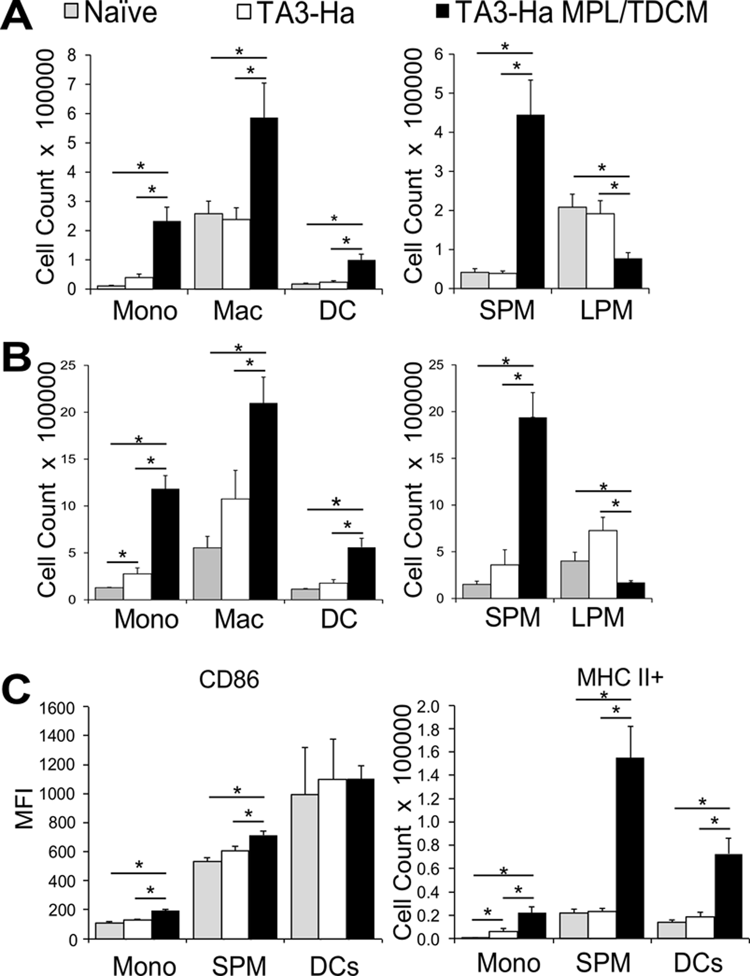
Figure 2. Myeloid-derived leukocyte populations are increased in the peritoneal cavity post MPL/TDCM treatment.

(A-B) Inflammatory monocytes (CD11b+ Ly6G− CD11c− B220− Ly6C+ SSClow), macrophages (CD11b+ Ly6G− CD11c− B220− Ly6Clow SSClow) and dendritic cells (cDC2; CD11b+ Ly6G− CD11c+ B220−) in peritoneal cavities on day 1 (A) and day 3 (B) post TA3-Ha cell challenge ± MPL/TDCM treatment on d0. Small peritoneal macrophages (SPM; CD11bmidF4/80mid) and large peritoneal macrophages (LPM; CD11bhiF4/80hi) were further quantified. (C) CD86 expression level (MFI) and MHC II+ monocyte and SPM cell numbers on day 1 post-tumor challenge. Values represent means ± SEM. Asterisks (*) indicate significant differences between indicated groups (p<0.05; n=3–5 mice/group.) (Mono: monocyte, Mac: macrophage, DC: dendritic cell, SPM: small peritoneal macrophage, LPM: large peritoneal macrophage). Results and statistical analysis for one of two representative experiments are shown.
