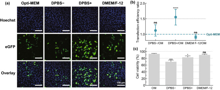
Fig. 3.
Screening of transfection buffers for mRNA transfection in HeLa cells. HeLa cells were transfected with eGFP-mRNA by VNB photoporation (0.3 µM mRNA; 8 × 10⁷ AuNPs mL−1; 3.6 J cm−2) using different transfection buffers: Opti-MEM (OM), DPBS−, DPBS+ or DMEM/F-12. a Representative confocal microscopy images of HeLa cells 24 h after transfection (Scale bar = 150 µm). b Transfection efficiency ratios for different buffers compared to OM, as measured by flow cytometry (n = 6). A one-way ANOVA with Dunnett’s multiple comparison test was performed to determine statistical differences between OM and the other buffers (ns = nonsignificant; ****p < .0001). c Cell viability 24 h post transfection, expressed relatively to the untreated control (n = 3). A one-way ANOVA with Dunnett’s multiple comparison test was performed to determine statistical differences between OM and the other buffers (ns = nonsignificant; *p < .05; ***p < .001)

