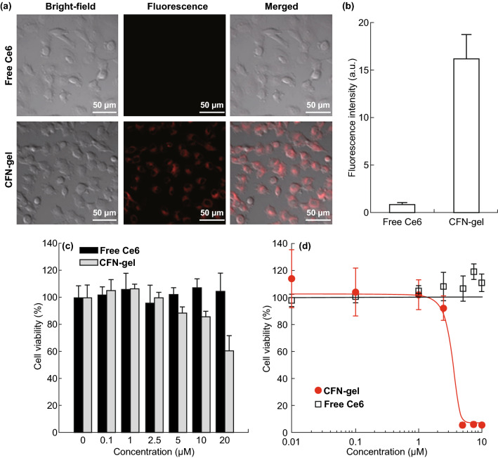
Fig. 5.
a NIR fluorescence images of free Ce6 and CFN-gel-treated HT1080 cancer cells. The HT1080 cancer cells were treated with free Ce6 or CFN-gel at a 2 µM Ce6 equivalent for 6 h. After washing the cells, NIR fluorescence images (λex = 633 nm, λem = 650, using a long-pass filter) were obtained. The red color indicates the fluorescence signals generated from Ce6, b quantitative analysis of fluorescence intensities in the free Ce6 and CFN-gel-treated HT1080 cells (n = 4), c in vitro dark cytotoxicity of the HT1080 cells after treatment with free Ce6 and CFN-gel at various concentrations, d in vitro phototoxicity of the HT1080 cells after treatment with free Ce6 and CFN-gel at various concentrations upon 670-nm light irradiation (50 mW cm−2, 10 J cm−2). The IC50 of the CFN-gel is 2.73 µM. (Color figure online)

