Fig. 1.
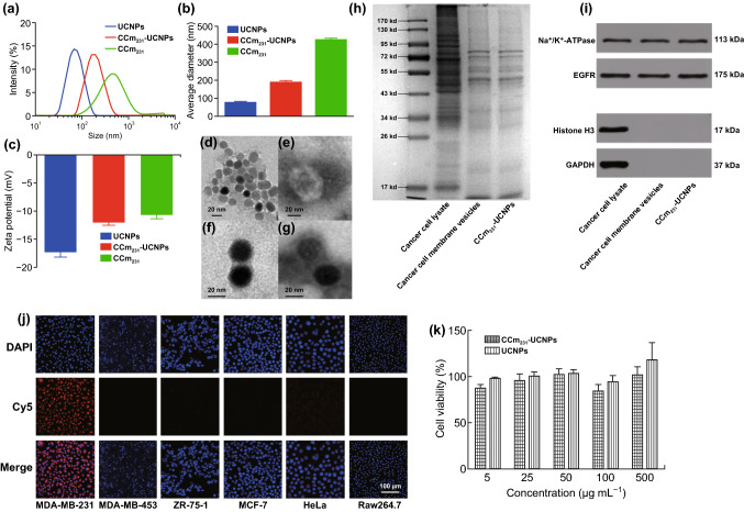
Physicochemical characterization of CCm231-UCNPs. a Size intensity curves, b hydrodynamic size, and c zeta potential of UCNPs, CCm231-UCNPs, and CCm231 measured by dynamic light scattering (DLS). TEM images of d UCNPs, e CCm231, and f, g CCm231-UCNPs. The TEM samples were negatively stained with phosphotungstic acid. The scale bars = 20 nm. h SDS-PAGE protein analysis of MDA-MB-231 cancer cell lysate, cancer cell membrane vesicles, and CCm231-UCNPs. Samples were run at equal protein concentration and stained with Coomassie Blue. i Western blotting analysis. Samples were run at equal protein concentration and immunostained against membrane markers including Na+/K+-ATPase, EGFR, histone H3 (a nuclear marker), and glyceraldehyde 3-phosphate dehydrogenase (a cytosolic marker). In vitro homologous-targeting ability and cytotoxicity of CCm231-UCNPs. j Confocal laser scanning microscopy (CLSM) images of various cells after incubation with CCm231-UCNPs. The scale bars = 100 μm. k Survival of MDA-MB-231 cells treated with different concentrations of CCm231-UCNPs and UCNPs. The data points represent mean ± SD (n = 3)

