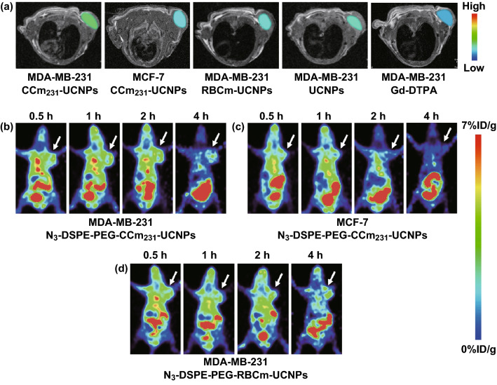
Fig. 3.
In vivo MR imaging. T1-weighted MR images acquired at 24 h after the injection of nanoparticles. a BALB/c nude tumor-bearing mice injected with Gd-DTPA or PBS containing CCm231-UCNPs, RBCm-UCNPs, UCNPs. The tumor sites are color-coded to better show the contrast-enhancing effects. In vivo PET imaging. Micro-PET static imaging was performed at 0.5, 1, 2, and 4 h after injection of Al[18F]F-L-NETA-DBCO. b MDA-MB-231 tumor-bearing mice injected with N3-DSPE-PEG-CCm231-UCNPs. c MCF-7 tumor-bearing mice injected with N3-DSPE-PEG-CCm231-UCNPs. d MDA-MB-231 tumor-bearing mice injected with N3-DSPE-PEG-RBCm-UCNPs

