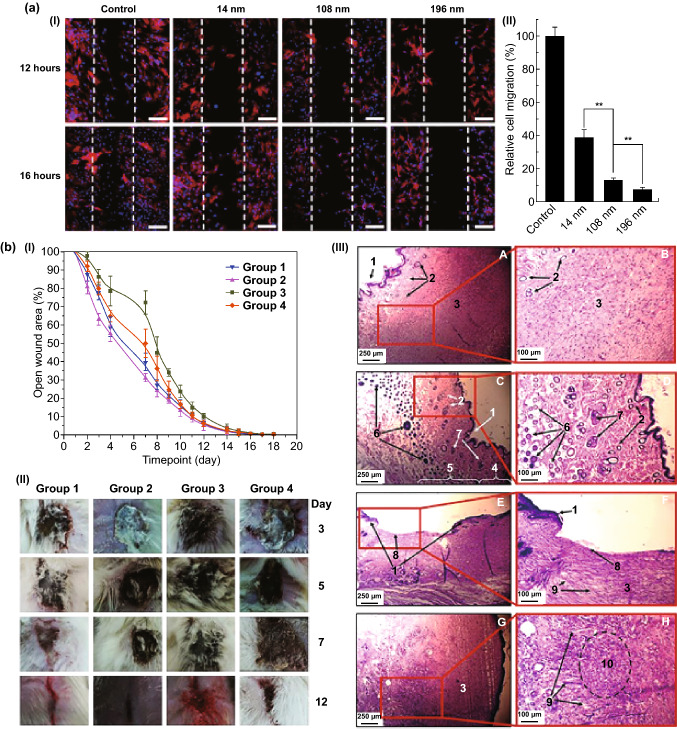
Fig. 10.
a Cellular migration of mesenchymal stem cells incubated with different sized TiO2 particles. Scale bars show 100 μm. Adapted from Ref. [208] with permission from the Dove Press Ltd. b The wound-healing process (I) macroscopically analyzed over the course of 19 days, (II) the representative wounds through the healing process for each group, and (III) the optical images of the healed skin from group 1 (A and B), group 2 (C and D), group 3 (E and F), and group 4 (G and H). Numbers indicate tissue structural elements: 1—epidermis; 2—sweat glands; 3—scar; 4—derma; 5—hypodermis; 6—hair follicles; 7—sebaceous glands; 8—de-epithelialized scar tissue; 9—scar vessels; 10—inflammatory infiltration in the scar. Adapted from Ref. [214] with permission from the Springer Nature

