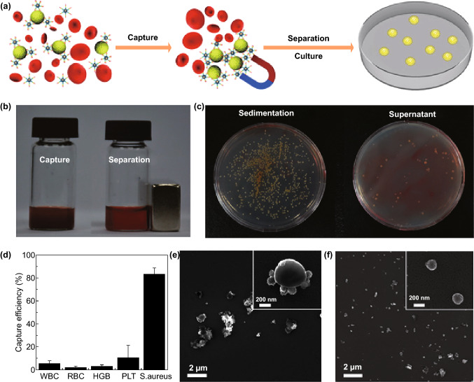
Fig. 9.
a The illustration represents the strategy for the identification and capture of pathogenic bacteria (S. aureus). b The photograph exhibits the capture (left) and separation (right) of bacteria with Fe3O4/TiO2 core–shell nanoparticles from an infected blood. c The number of colony-forming units in the re-cultured S. aureus from sediment and supernatant in agar plates after the separation. d Capture efficiency of different compounds in blood after being treated with AptS.aureus-Fe3O4@mTiO2. SEM images of the captured S. aureus (e) and non-captured E. coli (f) with the aptamer decorated nanoparticles.
Adapted from Ref. [28] with permission from the American Chemical Society

